Monday Poster Session
Category: Biliary/Pancreas
P1837 - A Rare Metastatic Presentation of Leiomyosarcoma Every Gastroenterologist Should Know
Monday, October 28, 2024
10:30 AM - 4:00 PM ET
Location: Exhibit Hall E

Has Audio

Astin Worden, MD
Mayo Clinic
Phoenix, AZ
Presenting Author(s)
Astin Worden, MD1, Danial Haris. Shaikh, MD2, Michelle Ann. Anderson, MD, MSc3
1Mayo Clinic, Phoenix, AZ; 2Mayo Clinic, Cave Creek, AZ; 3Mayo Clinic, Scottsdale, AZ
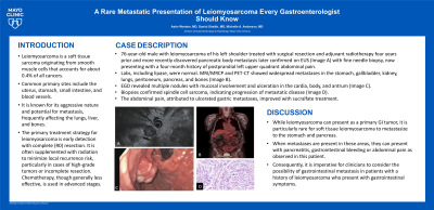
Introduction: Leiomyosarcoma is a rare soft tissue sarcoma originating from smooth muscle cells which accounts for about 0.4% of all cancers. Common primary sites include the uterus, stomach, small intestine, and blood vessels, particularly the inferior vena cava. It is known for its aggressive nature and potential for metastasis, frequently affecting the lungs, liver, and bones.
The primary treatment strategy for leiomyosarcoma is early detection with complete (R0) resection. It is often supplemented with radiation to minimize local recurrence risk, particularly in cases of high-grade tumors or incomplete resection. Chemotherapy, though generally less effective, is used in advanced stages. The 5-year survival rate for patients with metastatic leiomyosarcoma is approximately 13% overall, increasing to 20% following complete (R0) resection.
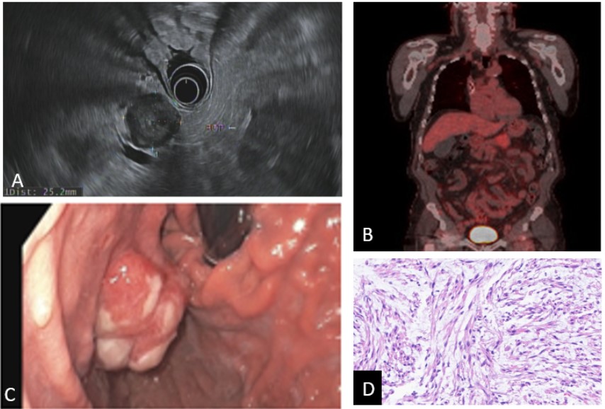
Case Description/Methods: We report a case involving a 76-year-old male previously diagnosed with leiomyosarcoma on his left shoulder, treated with surgical resection and adjuvant radiotherapy four years prior. Surveillance PET scan a year earlier revealed a 27 x 25 mm avid hypodense lesion in the pancreatic body, which was subsequently identified on EUS (Image A). Fine needle biopsy confirmed spindle cell sarcoma, consistent with recurrent leiomyosarcoma. The patient, initially treated with a gemcitabine/docetaxel regimen, was switched to oral pazopanib due to side effects.
Most recently, he presented with a four-month history of postprandial left upper quadrant abdominal pain. Laboratory tests, including lipase, were normal. MRI/MRCP and PET-CT showed widespread metastases in the stomach, gallbladder, kidney, lungs, peritoneum, pancreas, and bones (Image B). EGD revealed multiple nodules with mucosal involvement and ulceration in the cardia, body, and antrum (Image C). Biopsies confirmed spindle cell sarcoma, indicating progression of metastatic disease (Image D). The abdominal pain, attributed to ulcerated gastric metastasis, improved with sucralfate treatment.
Discussion: While leiomyosarcoma can present as a primary GI tumor, it is particularly rare for soft tissue leiomyosarcoma to metastasize to the stomach and pancreas. When metastases are present in these areas, they can present with pancreatitis, gastrointestinal bleeding or abdominal pain as observed in this patient. Consequently, it is imperative for clinicians to consider the possibility of gastrointestinal metastasis in patients with a history of leiomyosarcoma who present with gastrointestinal symptoms.

Disclosures:
Astin Worden, MD1, Danial Haris. Shaikh, MD2, Michelle Ann. Anderson, MD, MSc3. P1837 - A Rare Metastatic Presentation of Leiomyosarcoma Every Gastroenterologist Should Know, ACG 2024 Annual Scientific Meeting Abstracts. Philadelphia, PA: American College of Gastroenterology.
1Mayo Clinic, Phoenix, AZ; 2Mayo Clinic, Cave Creek, AZ; 3Mayo Clinic, Scottsdale, AZ
Introduction: Leiomyosarcoma is a rare soft tissue sarcoma originating from smooth muscle cells which accounts for about 0.4% of all cancers. Common primary sites include the uterus, stomach, small intestine, and blood vessels, particularly the inferior vena cava. It is known for its aggressive nature and potential for metastasis, frequently affecting the lungs, liver, and bones.
The primary treatment strategy for leiomyosarcoma is early detection with complete (R0) resection. It is often supplemented with radiation to minimize local recurrence risk, particularly in cases of high-grade tumors or incomplete resection. Chemotherapy, though generally less effective, is used in advanced stages. The 5-year survival rate for patients with metastatic leiomyosarcoma is approximately 13% overall, increasing to 20% following complete (R0) resection.
Case Description/Methods: We report a case involving a 76-year-old male previously diagnosed with leiomyosarcoma on his left shoulder, treated with surgical resection and adjuvant radiotherapy four years prior. Surveillance PET scan a year earlier revealed a 27 x 25 mm avid hypodense lesion in the pancreatic body, which was subsequently identified on EUS (Image A). Fine needle biopsy confirmed spindle cell sarcoma, consistent with recurrent leiomyosarcoma. The patient, initially treated with a gemcitabine/docetaxel regimen, was switched to oral pazopanib due to side effects.
Most recently, he presented with a four-month history of postprandial left upper quadrant abdominal pain. Laboratory tests, including lipase, were normal. MRI/MRCP and PET-CT showed widespread metastases in the stomach, gallbladder, kidney, lungs, peritoneum, pancreas, and bones (Image B). EGD revealed multiple nodules with mucosal involvement and ulceration in the cardia, body, and antrum (Image C). Biopsies confirmed spindle cell sarcoma, indicating progression of metastatic disease (Image D). The abdominal pain, attributed to ulcerated gastric metastasis, improved with sucralfate treatment.
Discussion: While leiomyosarcoma can present as a primary GI tumor, it is particularly rare for soft tissue leiomyosarcoma to metastasize to the stomach and pancreas. When metastases are present in these areas, they can present with pancreatitis, gastrointestinal bleeding or abdominal pain as observed in this patient. Consequently, it is imperative for clinicians to consider the possibility of gastrointestinal metastasis in patients with a history of leiomyosarcoma who present with gastrointestinal symptoms.

Figure: A) EUS showing hypoechoic homogenous pancreatic body mass B) PET CT with multiple tracer avid lesions C) Endoscopic image of gastric nodule with overlying ulceration D) Core needle section showing spindled cells arranged in intersection fascicles with nuclear atypia.
Disclosures:
Astin Worden indicated no relevant financial relationships.
Danial Shaikh indicated no relevant financial relationships.
Michelle Anderson: Boston Scientific – Consultant.
Astin Worden, MD1, Danial Haris. Shaikh, MD2, Michelle Ann. Anderson, MD, MSc3. P1837 - A Rare Metastatic Presentation of Leiomyosarcoma Every Gastroenterologist Should Know, ACG 2024 Annual Scientific Meeting Abstracts. Philadelphia, PA: American College of Gastroenterology.
