Monday Poster Session
Category: Biliary/Pancreas
P1902 - Simultaneous Occurrence of Adenocarcinomas in the Stomach and Biliary Tree: A Rare Case Report
Monday, October 28, 2024
10:30 AM - 4:00 PM ET
Location: Exhibit Hall E

Has Audio
- FN
Faisal Nimri, MD
Henry Ford Hospital
Detroit, MI
Presenting Author(s)
Faisal Nimri, MD1, Taher Jamali, MD1, Rund Nimri, MD2, Tariq Hammad, MD1, Tobias Zuchelli, MD1, Sumit Singla, MD1
1Henry Ford Hospital, Detroit, MI; 2Jordan University of Science and Technology, Irbid, Irbid, Jordan
Introduction: Synchronous adenocarcinomas in stomach and biliary tree are extremely rare phenomenon, with very scarce literature. The factors leading to the development of synchronous primary cancers within the GI tract remain poorly understood, but genetic predisposition, environment and molecular alterations may play a role.
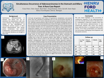
Case Description/Methods: A 66 year old man with history of hypertension, dyslipidemia, and prostate cancer was evaluated by hepatology clinic for new and persistent elevation of liver enzymes (LFTs) on routine check-up with ALT of 137 IU/L, AST of 60 IU/L, and ALP of 415 IU/L, while he had normal total bilirubin of 0.8 mg/dL, and INR 0.99. All LFTs were normal a year ago. He denied any associated symptoms. Further testing including viral hepatitis panel, autoimmune liver diseases, and hemochromatosis testing were negative. Liver US was normal. MRI with MRCP revealed a 2cm central lesion with left intrahepatic biliary ductal dilatation and questionable delayed enhancement. Advanced GI performed EGD which showed an ulcerated and friable tumor on the gastric antrum greater curvature concerning for malignancy and was biopsied. EUS to characterize the mass showed invasion into at least the muscularis propria. ERCP revealed a single moderate biliary stricture found in the left main hepatic duct. Exploration with cholangioscopy demonstrated focally abnormal epithelium concerning for malignancy, and was biopsied and brushing performed for FISH and cytology. The right hepatic duct was not involved though could be secondarily compressed. CBD and CHD were normal. A biliary sphincterotomy was performed. The stricture was dilated to 4 mm (with persistent waist) then stented with a 7 Fr by 12 cm straight plastic biliary stent. Pathological analysis of both gastric and left main hepatic duct stricture revealed moderately differentiated adenocarcinoma. FISH Bile Duct Malignancy panel showed evidence of trisomy 7 which has only a little evidence to suggest that trisomy 7 meets the criteria for a diagnosis of cholangiocarcinoma.
Discussion: This case report highlights a rare occurrence of simultaneous diagnosis of two adenocarcinomas, one in the stomach and the other in the biliary tree. It is challenging to determine whether these are two primary adenocarcinomas in two anatomically separate sites versus one primary lesion while the other is a metastatic lesion of that primary. Such a presentation is extremely rare and proposes challenging clinical diagnostic and therapeutic scenarios.

Note: The table for this abstract can be viewed in the ePoster Gallery section of the ACG 2024 ePoster Site or in The American Journal of Gastroenterology's abstract supplement issue, both of which will be available starting October 27, 2024.
Disclosures:
Faisal Nimri, MD1, Taher Jamali, MD1, Rund Nimri, MD2, Tariq Hammad, MD1, Tobias Zuchelli, MD1, Sumit Singla, MD1. P1902 - Simultaneous Occurrence of Adenocarcinomas in the Stomach and Biliary Tree: A Rare Case Report, ACG 2024 Annual Scientific Meeting Abstracts. Philadelphia, PA: American College of Gastroenterology.
1Henry Ford Hospital, Detroit, MI; 2Jordan University of Science and Technology, Irbid, Irbid, Jordan
Introduction: Synchronous adenocarcinomas in stomach and biliary tree are extremely rare phenomenon, with very scarce literature. The factors leading to the development of synchronous primary cancers within the GI tract remain poorly understood, but genetic predisposition, environment and molecular alterations may play a role.
Case Description/Methods: A 66 year old man with history of hypertension, dyslipidemia, and prostate cancer was evaluated by hepatology clinic for new and persistent elevation of liver enzymes (LFTs) on routine check-up with ALT of 137 IU/L, AST of 60 IU/L, and ALP of 415 IU/L, while he had normal total bilirubin of 0.8 mg/dL, and INR 0.99. All LFTs were normal a year ago. He denied any associated symptoms. Further testing including viral hepatitis panel, autoimmune liver diseases, and hemochromatosis testing were negative. Liver US was normal. MRI with MRCP revealed a 2cm central lesion with left intrahepatic biliary ductal dilatation and questionable delayed enhancement. Advanced GI performed EGD which showed an ulcerated and friable tumor on the gastric antrum greater curvature concerning for malignancy and was biopsied. EUS to characterize the mass showed invasion into at least the muscularis propria. ERCP revealed a single moderate biliary stricture found in the left main hepatic duct. Exploration with cholangioscopy demonstrated focally abnormal epithelium concerning for malignancy, and was biopsied and brushing performed for FISH and cytology. The right hepatic duct was not involved though could be secondarily compressed. CBD and CHD were normal. A biliary sphincterotomy was performed. The stricture was dilated to 4 mm (with persistent waist) then stented with a 7 Fr by 12 cm straight plastic biliary stent. Pathological analysis of both gastric and left main hepatic duct stricture revealed moderately differentiated adenocarcinoma. FISH Bile Duct Malignancy panel showed evidence of trisomy 7 which has only a little evidence to suggest that trisomy 7 meets the criteria for a diagnosis of cholangiocarcinoma.
Discussion: This case report highlights a rare occurrence of simultaneous diagnosis of two adenocarcinomas, one in the stomach and the other in the biliary tree. It is challenging to determine whether these are two primary adenocarcinomas in two anatomically separate sites versus one primary lesion while the other is a metastatic lesion of that primary. Such a presentation is extremely rare and proposes challenging clinical diagnostic and therapeutic scenarios.

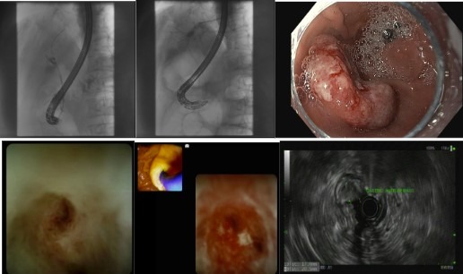
Figure: ERCP and cholangioscopy showing stricture resembling cholangiocarcinoma. Endoscopy showing gastric mass with EUS showing invasion to muscularis propria.
Note: The table for this abstract can be viewed in the ePoster Gallery section of the ACG 2024 ePoster Site or in The American Journal of Gastroenterology's abstract supplement issue, both of which will be available starting October 27, 2024.
Disclosures:
Faisal Nimri indicated no relevant financial relationships.
Taher Jamali indicated no relevant financial relationships.
Rund Nimri indicated no relevant financial relationships.
Tariq Hammad indicated no relevant financial relationships.
Tobias Zuchelli: Boston Scientific – Consultant.
Sumit Singla: Boston Scientific – Consultant.
Faisal Nimri, MD1, Taher Jamali, MD1, Rund Nimri, MD2, Tariq Hammad, MD1, Tobias Zuchelli, MD1, Sumit Singla, MD1. P1902 - Simultaneous Occurrence of Adenocarcinomas in the Stomach and Biliary Tree: A Rare Case Report, ACG 2024 Annual Scientific Meeting Abstracts. Philadelphia, PA: American College of Gastroenterology.
