Monday Poster Session
Category: Biliary/Pancreas
P1909 - The First Reported Case of a Pseudosarcomatous Stromal Tumor: A Unique Benign Ampullary Tumor
Monday, October 28, 2024
10:30 AM - 4:00 PM ET
Location: Exhibit Hall E

Has Audio
- JJ
Jacob C. Jones, DO
Brooke Army Medical Center
San Antonio, TX
Presenting Author(s)
Award: Presidential Poster Award
Penelope Quiles, 1, Jacob C. Jones, DO2, John G. Quiles, MD2, Jerome C. Edelson, MD2
1Washington and Lee University, San Antonio, TX; 2Brooke Army Medical Center, San Antonio, TX
Introduction: Pseudosarcomatous stromal proliferation is a rare, benign lesion usually found in submucosal stroma of the bladder, with few reports in the GI tract. Treatments include surgical resection, endoscopic resection, laser ablation, photodynamic therapy, and/or chemotherapy. Here, we present a case of pseudosarcomatous stromal proliferation involving the ampullary and duodenal mucosa.
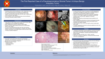
Case Description/Methods: A 61-year-old male with a history of prostate and carotid paraganglioma and metabolic associated liver disease was incidentally found to have a duodenal mass on MRI obtained to assess liver stiffness. The patient was asymptomatic and was referred for endoscopic visualization. A 20 mm x 17 mm, friable, fungating, and ulcerated non-obstructing ampullary mass was discovered, suggestive of ampullary adenocarcinoma. Biopsies were obtained and demonstrated reactive epithelial with stromal changes. Multidisciplinary discussion was performed including Surgical Oncology, with recommendations for EUS with FNB, which again demonstrated stromal proliferation and reactive changes. Labs to include CA 19-9, CA-125, CEA, IgG4, and liver function tests were normal. The patient subsequently underwent endoscopic papillectomy which was successful with R0 resection. Pathology demonstrated substantial fibroblastic/myofibroblastic stromal proliferation involving the ampullary and duodenal mucosa with marked reactive epithelial changes and gastric mucin cell metaplasia, consistent with pseudosarcomatous stromal changes. There was no evidence of any malignancy, including sarcomatoid prostate carcinoma or paraganglioma.
Discussion: This is the first known case of pseudosarcomatous stromal proliferation involving the ampullary and duodenal mucosa. Pseudosarcomatous stromal proliferation is typically found in the urogenital tract, though few have been reported within GI tract. Given the patients benign presentation, surgical management was deferred in favor of endoscopic papillectomy. This pseudosarcomatous stromal proliferation and its treatment is unprecedented, therefore, should serve as an example for similar cases.

Disclosures:
Penelope Quiles, 1, Jacob C. Jones, DO2, John G. Quiles, MD2, Jerome C. Edelson, MD2. P1909 - The First Reported Case of a Pseudosarcomatous Stromal Tumor: A Unique Benign Ampullary Tumor, ACG 2024 Annual Scientific Meeting Abstracts. Philadelphia, PA: American College of Gastroenterology.
Penelope Quiles, 1, Jacob C. Jones, DO2, John G. Quiles, MD2, Jerome C. Edelson, MD2
1Washington and Lee University, San Antonio, TX; 2Brooke Army Medical Center, San Antonio, TX
Introduction: Pseudosarcomatous stromal proliferation is a rare, benign lesion usually found in submucosal stroma of the bladder, with few reports in the GI tract. Treatments include surgical resection, endoscopic resection, laser ablation, photodynamic therapy, and/or chemotherapy. Here, we present a case of pseudosarcomatous stromal proliferation involving the ampullary and duodenal mucosa.
Case Description/Methods: A 61-year-old male with a history of prostate and carotid paraganglioma and metabolic associated liver disease was incidentally found to have a duodenal mass on MRI obtained to assess liver stiffness. The patient was asymptomatic and was referred for endoscopic visualization. A 20 mm x 17 mm, friable, fungating, and ulcerated non-obstructing ampullary mass was discovered, suggestive of ampullary adenocarcinoma. Biopsies were obtained and demonstrated reactive epithelial with stromal changes. Multidisciplinary discussion was performed including Surgical Oncology, with recommendations for EUS with FNB, which again demonstrated stromal proliferation and reactive changes. Labs to include CA 19-9, CA-125, CEA, IgG4, and liver function tests were normal. The patient subsequently underwent endoscopic papillectomy which was successful with R0 resection. Pathology demonstrated substantial fibroblastic/myofibroblastic stromal proliferation involving the ampullary and duodenal mucosa with marked reactive epithelial changes and gastric mucin cell metaplasia, consistent with pseudosarcomatous stromal changes. There was no evidence of any malignancy, including sarcomatoid prostate carcinoma or paraganglioma.
Discussion: This is the first known case of pseudosarcomatous stromal proliferation involving the ampullary and duodenal mucosa. Pseudosarcomatous stromal proliferation is typically found in the urogenital tract, though few have been reported within GI tract. Given the patients benign presentation, surgical management was deferred in favor of endoscopic papillectomy. This pseudosarcomatous stromal proliferation and its treatment is unprecedented, therefore, should serve as an example for similar cases.

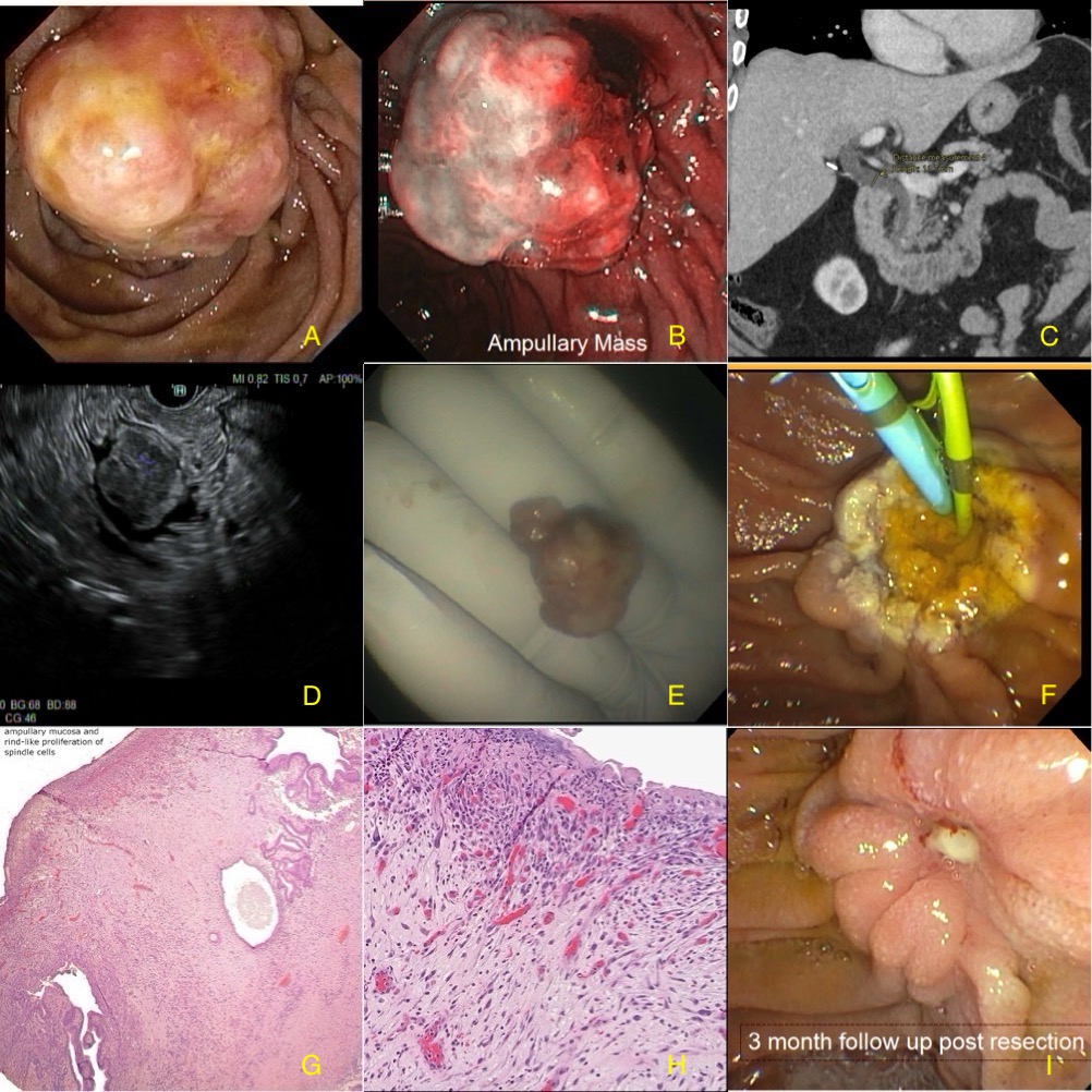
Figure: Image A: Ampullary duct lesion
Image B: Ampullary duct lesion with narrow band imaging
Image C: CT imaging of ampullary duct lesion
Image D: EUS of ampullary duct lesion
Image E: Post endoscopic resection
Image F: Stent placed in pancreatic duct and bile duct
Image G: Ampullary mucosa and rind-like proliferation of spindle cells
Image H: High-power view
Image I: 3 month follow up post resection
Disclosures:
Penelope Quiles indicated no relevant financial relationships.
Jacob Jones indicated no relevant financial relationships.
John Quiles indicated no relevant financial relationships.
Jerome Edelson indicated no relevant financial relationships.
Penelope Quiles, 1, Jacob C. Jones, DO2, John G. Quiles, MD2, Jerome C. Edelson, MD2. P1909 - The First Reported Case of a Pseudosarcomatous Stromal Tumor: A Unique Benign Ampullary Tumor, ACG 2024 Annual Scientific Meeting Abstracts. Philadelphia, PA: American College of Gastroenterology.

