Monday Poster Session
Category: Colon
P1983 - Rectal Ulcer and Fistula Development in a Cancer Patient on Immune Checkpoint Inhibitor Immunotherapy: A Case Report
Monday, October 28, 2024
10:30 AM - 4:00 PM ET
Location: Exhibit Hall E

Has Audio

Christine Catinis, MD
University of Texas at Houston
Houston, TX
Presenting Author(s)
Christine Catinis, MD1, Nitish Mittal, MD2, Parvir Aujla, MD2, Yinghong Wang, MD, PhD3
1University of Texas at Houston, Houston, TX; 2University of Texas Health, McGovern Medical School, Houston, TX; 3University of Texas MD Anderson Cancer Center, Houston, TX
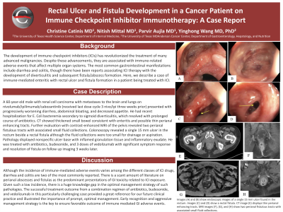
Introduction: Immune checkpoint inhibitors (ICIs) are associated with various immune-related adverse events. The most common gastrointestinal manifestations include diarrhea and colitis, though there have been reports associating ICI therapy with the development of diverticulitis and subsequent fistula/abscess formation. Here, we describe a case of immune-mediated enteritis with rectal ulcer and fistula formation in a patient being treated with immunotherapy.
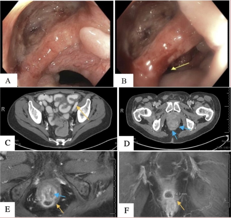
Case Description/Methods: A 64-year-old male with metastatic renal cell carcinoma presented to the emergency room with progressively worsening diarrhea, abdominal bloating, and decreased appetite. He was receiving treatment with nivolumab/ipilimumab and cabozantinib. Upon arrival to the hospital, Cabozantinib was held but his symptoms continued to worsen. Initial testing revealed a negative enteric stool pathogen panel, negative C. difficile PCR/antigen, positive lactoferrin, and calprotectin of 955 mcg/gm. CT of the abdomen and pelvis showed interval development of immune-mediated enteritis and possible thin perianal enhancing tracts. Further evaluation with contrast-enhanced MRI of the pelvis confirmed the presence of two perianal fistulous tracts with associated small fluid collections.
The patient was started on antibiotics and underwent colonoscopy, which revealed a single 15 mm rectal ulcer distorting the anal canal and distal rectum alongside a fistula opening. Biopsies were obtained and showed a nonspecific ulcer base with inflamed granulation tissue and inflammatory exudate. Given the patient’s persistent diarrhea secondary to presumed immune-mediated enteritis, he was started on budesonide and vedolizumab with significant symptom improvement. After his second dose of vedolizumab, his diarrheal symptoms completely resolved. Repeat imaging revealed resolution of the enteritis and rectal fistula. In addition, a repeat calprotectin level was < 50 mcg/gm, therefore, treatment with budesonide was discontinued. The patient received a total of three doses of vedolizumab. Cabozantinib was re-started at a lower dose, together with nivolumab monotherapy.
Discussion: Perianal abscesses and fistulas as the predominant presentations of GI toxicity post ICI exposure have been rarely reported, and thus treatment is predominantly based on anecdotal evidence. The successful treatment outcome from a combination regimen of antibiotics, budesonide, and vedolizumab in this particularly challenging case provides a great reference for our future clinical practice.

Disclosures:
Christine Catinis, MD1, Nitish Mittal, MD2, Parvir Aujla, MD2, Yinghong Wang, MD, PhD3. P1983 - Rectal Ulcer and Fistula Development in a Cancer Patient on Immune Checkpoint Inhibitor Immunotherapy: A Case Report, ACG 2024 Annual Scientific Meeting Abstracts. Philadelphia, PA: American College of Gastroenterology.
1University of Texas at Houston, Houston, TX; 2University of Texas Health, McGovern Medical School, Houston, TX; 3University of Texas MD Anderson Cancer Center, Houston, TX
Introduction: Immune checkpoint inhibitors (ICIs) are associated with various immune-related adverse events. The most common gastrointestinal manifestations include diarrhea and colitis, though there have been reports associating ICI therapy with the development of diverticulitis and subsequent fistula/abscess formation. Here, we describe a case of immune-mediated enteritis with rectal ulcer and fistula formation in a patient being treated with immunotherapy.
Case Description/Methods: A 64-year-old male with metastatic renal cell carcinoma presented to the emergency room with progressively worsening diarrhea, abdominal bloating, and decreased appetite. He was receiving treatment with nivolumab/ipilimumab and cabozantinib. Upon arrival to the hospital, Cabozantinib was held but his symptoms continued to worsen. Initial testing revealed a negative enteric stool pathogen panel, negative C. difficile PCR/antigen, positive lactoferrin, and calprotectin of 955 mcg/gm. CT of the abdomen and pelvis showed interval development of immune-mediated enteritis and possible thin perianal enhancing tracts. Further evaluation with contrast-enhanced MRI of the pelvis confirmed the presence of two perianal fistulous tracts with associated small fluid collections.
The patient was started on antibiotics and underwent colonoscopy, which revealed a single 15 mm rectal ulcer distorting the anal canal and distal rectum alongside a fistula opening. Biopsies were obtained and showed a nonspecific ulcer base with inflamed granulation tissue and inflammatory exudate. Given the patient’s persistent diarrhea secondary to presumed immune-mediated enteritis, he was started on budesonide and vedolizumab with significant symptom improvement. After his second dose of vedolizumab, his diarrheal symptoms completely resolved. Repeat imaging revealed resolution of the enteritis and rectal fistula. In addition, a repeat calprotectin level was < 50 mcg/gm, therefore, treatment with budesonide was discontinued. The patient received a total of three doses of vedolizumab. Cabozantinib was re-started at a lower dose, together with nivolumab monotherapy.
Discussion: Perianal abscesses and fistulas as the predominant presentations of GI toxicity post ICI exposure have been rarely reported, and thus treatment is predominantly based on anecdotal evidence. The successful treatment outcome from a combination regimen of antibiotics, budesonide, and vedolizumab in this particularly challenging case provides a great reference for our future clinical practice.

Figure: Figure 1: Colonoscopy images showing (A) a single 15 mm rectal ulcer and (B) a rectal fistula opening (yellow arrow). Contrast enhanced CT images (coronal) showing (C) thickened wall of the small bowel with peri-enteric stranding (yellow arrow), suggestive of immunotherapy related enteritis, and (D) thin perianal enhancing tracts (blue arrows). Contrast enhanced MRI images (coronal) showing (E) a perianal fistulous tract (blue arrow) associated with a 0.9 cm fluid collection (yellow arrow) and (F) a 1.4 cm fluid collection (yellow arrow).
Disclosures:
Christine Catinis indicated no relevant financial relationships.
Nitish Mittal indicated no relevant financial relationships.
Parvir Aujla indicated no relevant financial relationships.
Yinghong Wang: AzurRx – Consultant. Ilyapharma – Consultant. IOTA – Consultant. Sorriso – Consultant. Tillotts – Consultant.
Christine Catinis, MD1, Nitish Mittal, MD2, Parvir Aujla, MD2, Yinghong Wang, MD, PhD3. P1983 - Rectal Ulcer and Fistula Development in a Cancer Patient on Immune Checkpoint Inhibitor Immunotherapy: A Case Report, ACG 2024 Annual Scientific Meeting Abstracts. Philadelphia, PA: American College of Gastroenterology.
