Monday Poster Session
Category: Colon
P1988 - A Rare Presentation of Cytomegalovirus (CMV) Colitis as a Colonic Mass
Monday, October 28, 2024
10:30 AM - 4:00 PM ET
Location: Exhibit Hall E

Has Audio

Mohammed Abusuliman, MD
Henry Ford Health
Detroit, MI
Presenting Author(s)
Mohammed Abusuliman, MD1, Amr Abusuliman, MD2, Abdulmalik Saleem, MD1, Ahmad Alomari, MD3, Hazem Abosheaishaa, MD4, Faisal Nimri, MD3, Taher Jamali, MD3, Najwa El-Nachef, MD1
1Henry Ford Health, Detroit, MI; 2Tanta University, Qutour, Al Gharbiyah, Egypt; 3Henry Ford Hospital, Detroit, MI; 4Icahn School of Medicine at Mount Sinai, Queens, NY
Introduction: Cytomegalovirus (CMV) infection affects the gastrointestinal tract in both immunocompromised and immunocompetent individuals. While commonly manifesting as ulcerative lesions, instances of CMV-induced mass lesions in the gastrointestinal tract are rare, and have only been described in a handful of cases. We herein report a rare presentation of CMV as a discrete colonic mass in a patient presenting with hematochezia.
Case Description/Methods: a 54-year-old female with end state renal disease on hemodialysis, hypertension, and sarcoidosis (on azathioprine), presented to the emergency room (ER) with rectal bleeding ongoing for a few months.
A month prior she was hospitalized for diarrhea and abdominal pain. Abdominal computed tomography (CT) at the time was concerning for ischemic vs infectious colitis. She was scheduled for an outpatient colonoscopy.
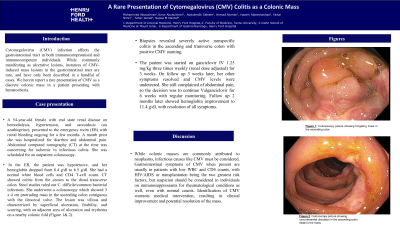
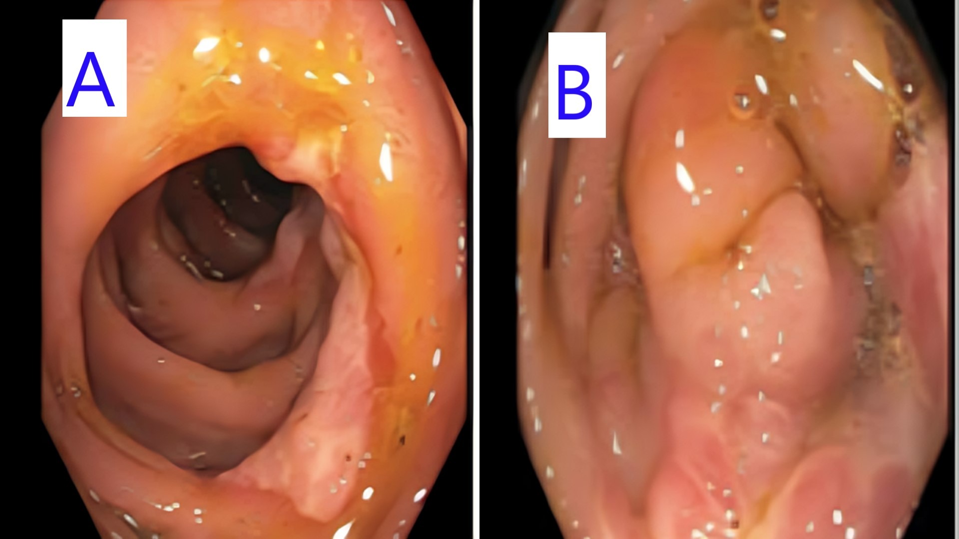
In the ER, the patient was hypotensive, and her hemoglobin dropped from 8.4 g/dl to 6.5 g/dl. She had a normal white blood cells and CD4 T-cell count. CT showed colitis from the cecum to the distal transverse colon. Stool studies ruled out C. difficile/common bacterial infections. She underwent a colonoscopy which showed 3 x 4 cm protruding mass in the ascending colon contiguous with the ileocecal valve. The lesion was villous and characterized by superficial ulceration, friability, and scarring, with an adjacent area of ulceration and erythema on a nearby colonic fold (Figure 1). Biopsies revealed severely active nonspecific colitis in the ascending and transverse colon with positive CMV staining.
The patient was started on ganciclovir IV 1.25 mg/kg three times weekly (renal dose adjusted) for 3 weeks. On follow up 3 weeks later, her other symptoms resolved and CMV levels were undetected. She still complained of abdominal pain, so the decision was to continue Valganciclovir for 6 weeks with regular monitoring. Follow up 2 months later showed hemoglobin improvement to 11.4 g/dl, with resolution of all symptoms.
Discussion: While colonic masses are commonly attributed to neoplasms, infectious causes like CMV must be considered. Gastrointestinal symptoms of CMV when present are usually in patients with low WBC and CD4 counts, with HIV/AIDS or transplantation being the two greatest risk factors, but suspicion should be considered in individuals on immunosuppressants for rheumatological conditions as well, even with normal counts. Identification of CMV warrants medical intervention, resulting in clinical improvement and potential resolution of the mass.

Disclosures:
Mohammed Abusuliman, MD1, Amr Abusuliman, MD2, Abdulmalik Saleem, MD1, Ahmad Alomari, MD3, Hazem Abosheaishaa, MD4, Faisal Nimri, MD3, Taher Jamali, MD3, Najwa El-Nachef, MD1. P1988 - A Rare Presentation of Cytomegalovirus (CMV) Colitis as a Colonic Mass, ACG 2024 Annual Scientific Meeting Abstracts. Philadelphia, PA: American College of Gastroenterology.
1Henry Ford Health, Detroit, MI; 2Tanta University, Qutour, Al Gharbiyah, Egypt; 3Henry Ford Hospital, Detroit, MI; 4Icahn School of Medicine at Mount Sinai, Queens, NY
Introduction: Cytomegalovirus (CMV) infection affects the gastrointestinal tract in both immunocompromised and immunocompetent individuals. While commonly manifesting as ulcerative lesions, instances of CMV-induced mass lesions in the gastrointestinal tract are rare, and have only been described in a handful of cases. We herein report a rare presentation of CMV as a discrete colonic mass in a patient presenting with hematochezia.
Case Description/Methods: a 54-year-old female with end state renal disease on hemodialysis, hypertension, and sarcoidosis (on azathioprine), presented to the emergency room (ER) with rectal bleeding ongoing for a few months.
A month prior she was hospitalized for diarrhea and abdominal pain. Abdominal computed tomography (CT) at the time was concerning for ischemic vs infectious colitis. She was scheduled for an outpatient colonoscopy.
In the ER, the patient was hypotensive, and her hemoglobin dropped from 8.4 g/dl to 6.5 g/dl. She had a normal white blood cells and CD4 T-cell count. CT showed colitis from the cecum to the distal transverse colon. Stool studies ruled out C. difficile/common bacterial infections. She underwent a colonoscopy which showed 3 x 4 cm protruding mass in the ascending colon contiguous with the ileocecal valve. The lesion was villous and characterized by superficial ulceration, friability, and scarring, with an adjacent area of ulceration and erythema on a nearby colonic fold (Figure 1). Biopsies revealed severely active nonspecific colitis in the ascending and transverse colon with positive CMV staining.
The patient was started on ganciclovir IV 1.25 mg/kg three times weekly (renal dose adjusted) for 3 weeks. On follow up 3 weeks later, her other symptoms resolved and CMV levels were undetected. She still complained of abdominal pain, so the decision was to continue Valganciclovir for 6 weeks with regular monitoring. Follow up 2 months later showed hemoglobin improvement to 11.4 g/dl, with resolution of all symptoms.
Discussion: While colonic masses are commonly attributed to neoplasms, infectious causes like CMV must be considered. Gastrointestinal symptoms of CMV when present are usually in patients with low WBC and CD4 counts, with HIV/AIDS or transplantation being the two greatest risk factors, but suspicion should be considered in individuals on immunosuppressants for rheumatological conditions as well, even with normal counts. Identification of CMV warrants medical intervention, resulting in clinical improvement and potential resolution of the mass.

Figure: Figure 1: Colonscopy picture showing circumferential ulceration in the ascending colon distal to the mass (Panel A), colonscopy picture showing fungating mass in the ascending colon (Panel B)
Disclosures:
Mohammed Abusuliman indicated no relevant financial relationships.
Amr Abusuliman indicated no relevant financial relationships.
Abdulmalik Saleem indicated no relevant financial relationships.
Ahmad Alomari indicated no relevant financial relationships.
Hazem Abosheaishaa indicated no relevant financial relationships.
Faisal Nimri indicated no relevant financial relationships.
Taher Jamali indicated no relevant financial relationships.
Najwa El-Nachef: Abbvie – Grant/Research Support. Ferring Pharmaceuticals – Advisor or Review Panel Member.
Mohammed Abusuliman, MD1, Amr Abusuliman, MD2, Abdulmalik Saleem, MD1, Ahmad Alomari, MD3, Hazem Abosheaishaa, MD4, Faisal Nimri, MD3, Taher Jamali, MD3, Najwa El-Nachef, MD1. P1988 - A Rare Presentation of Cytomegalovirus (CMV) Colitis as a Colonic Mass, ACG 2024 Annual Scientific Meeting Abstracts. Philadelphia, PA: American College of Gastroenterology.
