Monday Poster Session
Category: Colon
P2033 - A Rare Case of Mucosal Schwann Cell Hamartoma
Monday, October 28, 2024
10:30 AM - 4:00 PM ET
Location: Exhibit Hall E


Ahmed Nadeem, MD
Cleveland Clinic Foundation
Cleveland, OH
Presenting Author(s)
Ahmed Nadeem, MD, Zehra Naseem, MD, Fatima Zehra Shah, MD, Natalie Farha, MD, Mohamed El Hag, MD, MS, Ari Garber, MD, MS
Cleveland Clinic Foundation, Cleveland, OH
Introduction: Mucosal Schwann Cell Hamartomas (MSCH) are a set of intramucosal tumors, mesenchymal in origin, found rarely in the gastrointestinal (GI) tract. They most commonly occur in the large intestine, however, there have been reports of their presence in other areas of the GI tract. MSCHs are usually not associated with any inherited familial syndromes. It is important to differentiate GI stromal tumors (GISTs) from GI Schwann cell hamartomas as the former have an increased risk of malignant transformation. Here, we present a case of MSCH in a patient with a family history of colorectal cancer identified during surveillance colonoscopy.
Case Description/Methods: A 50-year-old female with a past medical history of cervical dysplasia, hypercholesterolemia, obesity, and mild intermittent asthma presented to her outpatient primary clinic for her annual physical exam. She had no active complaints. She denied weight loss, constipation, diarrhea, abdominal pain, nausea, or vomiting. Her physical exam was unrevealing without any abdominal distention or skin lesions. Her family history was positive for colorectal cancer in her maternal grandmother in her late 40s. She had a routine screening colonoscopy in 2018 which revealed a 5 mm sessile polyp (biopsy: serrated polyp) in the cecum and sigmoid diverticulosis.
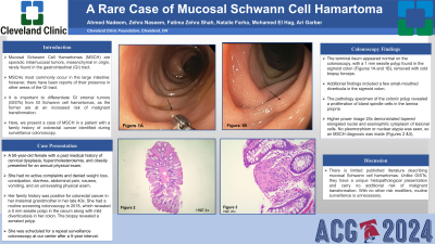
She was scheduled for a surveillance colonoscopy at our center after 6 years. A colonoscopy revealed a 1 mm sessile polyp in the sigmoid colon (Figures 1A and 1B), sigmoid diverticulosis, and small internal hemorrhoids found on retroflexion. The polyp was removed with a cold biopsy forceps. The pathology specimen of the colonic polyp revealed a proliferation of bland spindle cells in the lamina propria. A higher power image at 20x demonstrated tapered elongated nuclei and eosinophilic cytoplasm of lesional cells. No pleomorphism or nuclear atypia was seen. A diagnosis of a mucosal Schwann cell hamartoma was made (Figures 2 &3).
Discussion: There is limited published literature describing mucosal Schwann cell hamartomas. Unlike GISTs, they have a unique histopathological presentation and carry no additional risk of malignant transformation. With no other risk modifiers, routine surveillance is unnecessary.

Disclosures:
Ahmed Nadeem, MD, Zehra Naseem, MD, Fatima Zehra Shah, MD, Natalie Farha, MD, Mohamed El Hag, MD, MS, Ari Garber, MD, MS. P2033 - A Rare Case of Mucosal Schwann Cell Hamartoma, ACG 2024 Annual Scientific Meeting Abstracts. Philadelphia, PA: American College of Gastroenterology.
Cleveland Clinic Foundation, Cleveland, OH
Introduction: Mucosal Schwann Cell Hamartomas (MSCH) are a set of intramucosal tumors, mesenchymal in origin, found rarely in the gastrointestinal (GI) tract. They most commonly occur in the large intestine, however, there have been reports of their presence in other areas of the GI tract. MSCHs are usually not associated with any inherited familial syndromes. It is important to differentiate GI stromal tumors (GISTs) from GI Schwann cell hamartomas as the former have an increased risk of malignant transformation. Here, we present a case of MSCH in a patient with a family history of colorectal cancer identified during surveillance colonoscopy.
Case Description/Methods: A 50-year-old female with a past medical history of cervical dysplasia, hypercholesterolemia, obesity, and mild intermittent asthma presented to her outpatient primary clinic for her annual physical exam. She had no active complaints. She denied weight loss, constipation, diarrhea, abdominal pain, nausea, or vomiting. Her physical exam was unrevealing without any abdominal distention or skin lesions. Her family history was positive for colorectal cancer in her maternal grandmother in her late 40s. She had a routine screening colonoscopy in 2018 which revealed a 5 mm sessile polyp (biopsy: serrated polyp) in the cecum and sigmoid diverticulosis.
She was scheduled for a surveillance colonoscopy at our center after 6 years. A colonoscopy revealed a 1 mm sessile polyp in the sigmoid colon (Figures 1A and 1B), sigmoid diverticulosis, and small internal hemorrhoids found on retroflexion. The polyp was removed with a cold biopsy forceps. The pathology specimen of the colonic polyp revealed a proliferation of bland spindle cells in the lamina propria. A higher power image at 20x demonstrated tapered elongated nuclei and eosinophilic cytoplasm of lesional cells. No pleomorphism or nuclear atypia was seen. A diagnosis of a mucosal Schwann cell hamartoma was made (Figures 2 &3).
Discussion: There is limited published literature describing mucosal Schwann cell hamartomas. Unlike GISTs, they have a unique histopathological presentation and carry no additional risk of malignant transformation. With no other risk modifiers, routine surveillance is unnecessary.

Figure: Figure 1A and 1B- Sessile Polyp in sigmoid colon identified during colonoscopy
Figure 2- Biopsy of colon polyp with a proliferation of bland spindle cells in the lamina propria (4x)
Figure 3- Higher power image at 20x demonstrates tapered elongated nuclei and eosinophilic cytoplasm of lesional cells. No pleomorphism or nuclear atypia is seen.
Figure 2- Biopsy of colon polyp with a proliferation of bland spindle cells in the lamina propria (4x)
Figure 3- Higher power image at 20x demonstrates tapered elongated nuclei and eosinophilic cytoplasm of lesional cells. No pleomorphism or nuclear atypia is seen.
Disclosures:
Ahmed Nadeem indicated no relevant financial relationships.
Zehra Naseem indicated no relevant financial relationships.
Fatima Zehra Shah indicated no relevant financial relationships.
Natalie Farha indicated no relevant financial relationships.
Mohamed El Hag indicated no relevant financial relationships.
Ari Garber indicated no relevant financial relationships.
Ahmed Nadeem, MD, Zehra Naseem, MD, Fatima Zehra Shah, MD, Natalie Farha, MD, Mohamed El Hag, MD, MS, Ari Garber, MD, MS. P2033 - A Rare Case of Mucosal Schwann Cell Hamartoma, ACG 2024 Annual Scientific Meeting Abstracts. Philadelphia, PA: American College of Gastroenterology.
