Monday Poster Session
Category: Colon
P2040 - A Rare Case of Medullary Carcinoma of the Colon
Monday, October 28, 2024
10:30 AM - 4:00 PM ET
Location: Exhibit Hall E

Has Audio

Jose Martinez Perez, MD
University of California Davis Health
Sacramento, CA
Presenting Author(s)
Jose Martinez Perez, MD1, Kuldeep Tagore, MD1, Joseph Marsano, MD1, Omar Viramontes, MD2, Yesenia Ramos, MD1, Anupam Mitra, MBBS, MD1
1University of California Davis Health, Sacramento, CA; 2University of California Davis Health Graduate Medical Education, Sacramento, CA
Introduction: The majority of colorectal cancers (CRC) are adenocarcinomas. Medullary carcinoma is a rare subtype which represents 0.29% of all CRC. Medullary carcinoma is characterized by histologically distinct features such as undifferentiated high-grade cytology and prominent lymphocytic infiltration. It is frequently associated with microsatellite instability. It generally has better outcomes compared to poorly differentiated adenocarcinomas. Given its rarity, potential diagnostic and therapeutic approaches are topics of interest. In this case, we report this typically challenging pathologic diagnosis with an equally striking endoscopic golden appearance atypical for malignancy.
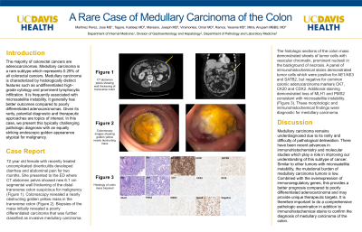
Case Description/Methods: 72 year old female with recently treated uncomplicated diverticulitis developed diarrhea and abdominal pain for two months. She presented to the ED where CT abdomen pelvis showed new 6.1 cm segmental wall thickening of the distal transverse colon suspicious for malignancy (figure a). Colonoscopy revealed a nearly obstructing golden yellow mass in the transverse colon (figure b). Biopsies of the mass initially revealed a poorly differentiated carcinoma that was further classified as invasive medullary carcinoma. The histologic sections of the colon mass demonstrated sheets of tumor cells with vesicular chromatin, prominent nucleoli in the background of necrosis. A panel of immunohistochemical stains demonstrated tumor cells which were positive for AE1/AE3 and SATB2, but negative for common colonic adenocarcinoma markers CK7, CK20 and CDX2. Additional staining demonstrated loss of MLH1 and PMS2 consistent with microsatellite instability. (fIgure c). These morphologic and immunohistochemical findings were diagnostic for medullary carcinoma.
Discussion: Medullary carcinoma remains underdiagnosed due to its rarity and difficulty of pathological delineation. There have been recent advances in immunohistochemistry and molecular studies which play a role in improving our understanding of this subtype of cancer. Similar to other tumors with microsatellite instability, the mutational burden of medullary carcinoma tumors is low. Combined with the overexpression of immunoregulatory genes, this provides a better prognosis compared to poorly differentiated adenocarcinoma and may provide unique therapeutic targets. It is therefore important to do a comprehensive pathologic examination in addition to immunohistochemical stains to confirm the diagnosis of medullary carcinoma of the colon.

Disclosures:
Jose Martinez Perez, MD1, Kuldeep Tagore, MD1, Joseph Marsano, MD1, Omar Viramontes, MD2, Yesenia Ramos, MD1, Anupam Mitra, MBBS, MD1. P2040 - A Rare Case of Medullary Carcinoma of the Colon, ACG 2024 Annual Scientific Meeting Abstracts. Philadelphia, PA: American College of Gastroenterology.
1University of California Davis Health, Sacramento, CA; 2University of California Davis Health Graduate Medical Education, Sacramento, CA
Introduction: The majority of colorectal cancers (CRC) are adenocarcinomas. Medullary carcinoma is a rare subtype which represents 0.29% of all CRC. Medullary carcinoma is characterized by histologically distinct features such as undifferentiated high-grade cytology and prominent lymphocytic infiltration. It is frequently associated with microsatellite instability. It generally has better outcomes compared to poorly differentiated adenocarcinomas. Given its rarity, potential diagnostic and therapeutic approaches are topics of interest. In this case, we report this typically challenging pathologic diagnosis with an equally striking endoscopic golden appearance atypical for malignancy.
Case Description/Methods: 72 year old female with recently treated uncomplicated diverticulitis developed diarrhea and abdominal pain for two months. She presented to the ED where CT abdomen pelvis showed new 6.1 cm segmental wall thickening of the distal transverse colon suspicious for malignancy (figure a). Colonoscopy revealed a nearly obstructing golden yellow mass in the transverse colon (figure b). Biopsies of the mass initially revealed a poorly differentiated carcinoma that was further classified as invasive medullary carcinoma. The histologic sections of the colon mass demonstrated sheets of tumor cells with vesicular chromatin, prominent nucleoli in the background of necrosis. A panel of immunohistochemical stains demonstrated tumor cells which were positive for AE1/AE3 and SATB2, but negative for common colonic adenocarcinoma markers CK7, CK20 and CDX2. Additional staining demonstrated loss of MLH1 and PMS2 consistent with microsatellite instability. (fIgure c). These morphologic and immunohistochemical findings were diagnostic for medullary carcinoma.
Discussion: Medullary carcinoma remains underdiagnosed due to its rarity and difficulty of pathological delineation. There have been recent advances in immunohistochemistry and molecular studies which play a role in improving our understanding of this subtype of cancer. Similar to other tumors with microsatellite instability, the mutational burden of medullary carcinoma tumors is low. Combined with the overexpression of immunoregulatory genes, this provides a better prognosis compared to poorly differentiated adenocarcinoma and may provide unique therapeutic targets. It is therefore important to do a comprehensive pathologic examination in addition to immunohistochemical stains to confirm the diagnosis of medullary carcinoma of the colon.

Figure: Figure a: CT abdomen pelvis showing thickening of distal transverse colon.
Figure b: Golden yellow mass seen on colonoscopy.
Figure c: Immunohistochemical stains of colon mass biopsy.
Figure b: Golden yellow mass seen on colonoscopy.
Figure c: Immunohistochemical stains of colon mass biopsy.
Disclosures:
Jose Martinez Perez indicated no relevant financial relationships.
Kuldeep Tagore indicated no relevant financial relationships.
Joseph Marsano indicated no relevant financial relationships.
Omar Viramontes indicated no relevant financial relationships.
Yesenia Ramos indicated no relevant financial relationships.
Anupam Mitra indicated no relevant financial relationships.
Jose Martinez Perez, MD1, Kuldeep Tagore, MD1, Joseph Marsano, MD1, Omar Viramontes, MD2, Yesenia Ramos, MD1, Anupam Mitra, MBBS, MD1. P2040 - A Rare Case of Medullary Carcinoma of the Colon, ACG 2024 Annual Scientific Meeting Abstracts. Philadelphia, PA: American College of Gastroenterology.
