Monday Poster Session
Category: Colon
P2053 - A Rare Case of a Cecal Inflammatory Fibroid Polyp
Monday, October 28, 2024
10:30 AM - 4:00 PM ET
Location: Exhibit Hall E

Has Audio

Dylan Nunn, MS
Heritage College of Osteopathic Medicine, Ohio University, OH
Presenting Author(s)
Dylan Nunn, MS1, Sara Yacyshyn, MD2, Drew Triplett, DO2
1Heritage College of Osteopathic Medicine, Ohio University, Dayton, OH; 2Wright State University, Dayton, OH
Introduction: Inflammatory fibroid polyps (IFPs) are rare benign mesenchymal tumors primarily found in the gastric antrum and small intestine, constituting less than 2% of all gastrointestinal (GI) polyps. They exhibit spindle cells arranged in an “onion-skin” pattern surrounding blood vessels, often accompanied by a submucosal inflammatory infiltrate. Several immunohistochemical stains aid in their differentiation from other spindle cell lesions. Two cases have shown a relationship between inflammatory bowel disease (IBD) and IFPs. Here, we present a case of incidental discovery of a large IFP in the cecum during endoscopic evaluation of new-onset Crohn’s disease.
Case Description/Methods: A 68-year-old female with a history of ulcerative colitis presented with abdominal pain. An abdominal CT scan revealed a thickened terminal ileum suggestive of Crohn's disease. Colonoscopy revealed thumbprinting and scarring throughout the colon, with multiple large inflammatory appearing polyps in the transverse and ascending colon. One large 4-cm mass-like lesion was identified in the cecum, which was lifted and resected in one piece by snare polypectomy and retrieved via a Roth net. Upon reinserting the colonoscope, a deformed cecum and inflammatory-appearing mucosa of the IC valve was revealed. Intubation of the terminal ileum showed an area of polypoid-like inflammation that was biopsied. The results of this biopsy confirmed the suspected diagnosis of Crohn’s disease. A panel of immunohistologic stains was utilized to evaluate the resected cecal specimen. These findings were consistent with an inflammatory fibroid polyp.
Discussion: IFPs are rare benign mesenchymal tumors found within the GI tract. IFPs of the colon are remarkably rare, with only 32 cases reported worldwide between 1952 and 2016. While they typically present asymptomatically, larger polyps have been reported to cause abdominal pain and obstruction. Our patient presented with abdominal pain but was also found to have new-onset Crohn's disease. Two case reports have been published regarding the simultaneous occurrence of active IBD and IFPs.
Treatment options for IFPs include surgical excision and endoscopic mucosal resection (EMR). There are no standardized treatment guidelines for IFPs; however, due to their benign nature and low recurrence rate, EMR is considered safe. For ileal IFPs less than 2cm in size, endoscopic resection was proven to be less invasive. We opted to remove this cecal IFP endoscopically based on similar parameters.

Disclosures:
Dylan Nunn, MS1, Sara Yacyshyn, MD2, Drew Triplett, DO2. P2053 - A Rare Case of a Cecal Inflammatory Fibroid Polyp, ACG 2024 Annual Scientific Meeting Abstracts. Philadelphia, PA: American College of Gastroenterology.
1Heritage College of Osteopathic Medicine, Ohio University, Dayton, OH; 2Wright State University, Dayton, OH
Introduction: Inflammatory fibroid polyps (IFPs) are rare benign mesenchymal tumors primarily found in the gastric antrum and small intestine, constituting less than 2% of all gastrointestinal (GI) polyps. They exhibit spindle cells arranged in an “onion-skin” pattern surrounding blood vessels, often accompanied by a submucosal inflammatory infiltrate. Several immunohistochemical stains aid in their differentiation from other spindle cell lesions. Two cases have shown a relationship between inflammatory bowel disease (IBD) and IFPs. Here, we present a case of incidental discovery of a large IFP in the cecum during endoscopic evaluation of new-onset Crohn’s disease.
Case Description/Methods: A 68-year-old female with a history of ulcerative colitis presented with abdominal pain. An abdominal CT scan revealed a thickened terminal ileum suggestive of Crohn's disease. Colonoscopy revealed thumbprinting and scarring throughout the colon, with multiple large inflammatory appearing polyps in the transverse and ascending colon. One large 4-cm mass-like lesion was identified in the cecum, which was lifted and resected in one piece by snare polypectomy and retrieved via a Roth net. Upon reinserting the colonoscope, a deformed cecum and inflammatory-appearing mucosa of the IC valve was revealed. Intubation of the terminal ileum showed an area of polypoid-like inflammation that was biopsied. The results of this biopsy confirmed the suspected diagnosis of Crohn’s disease. A panel of immunohistologic stains was utilized to evaluate the resected cecal specimen. These findings were consistent with an inflammatory fibroid polyp.
Discussion: IFPs are rare benign mesenchymal tumors found within the GI tract. IFPs of the colon are remarkably rare, with only 32 cases reported worldwide between 1952 and 2016. While they typically present asymptomatically, larger polyps have been reported to cause abdominal pain and obstruction. Our patient presented with abdominal pain but was also found to have new-onset Crohn's disease. Two case reports have been published regarding the simultaneous occurrence of active IBD and IFPs.
Treatment options for IFPs include surgical excision and endoscopic mucosal resection (EMR). There are no standardized treatment guidelines for IFPs; however, due to their benign nature and low recurrence rate, EMR is considered safe. For ileal IFPs less than 2cm in size, endoscopic resection was proven to be less invasive. We opted to remove this cecal IFP endoscopically based on similar parameters.

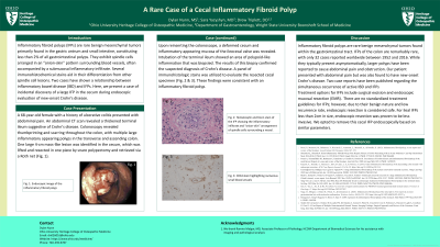
Figure: A) Endoscopic image of the IFP. B) Hematoxylin and eosin staining of the IFP showing an inflammatory infiltrate and “onion-skin” arrangement of spindle cells surrounding a vessel. C) CD34 stain highlighting numerous small blood vessels within the IFP.
Disclosures:
Dylan Nunn indicated no relevant financial relationships.
Sara Yacyshyn indicated no relevant financial relationships.
Drew Triplett indicated no relevant financial relationships.
Dylan Nunn, MS1, Sara Yacyshyn, MD2, Drew Triplett, DO2. P2053 - A Rare Case of a Cecal Inflammatory Fibroid Polyp, ACG 2024 Annual Scientific Meeting Abstracts. Philadelphia, PA: American College of Gastroenterology.
