Monday Poster Session
Category: Colon
P2093 - Colonic Rectal Schwannoma: A Case Report and Review of Literature
Monday, October 28, 2024
10:30 AM - 4:00 PM ET
Location: Exhibit Hall E

Has Audio

Matthew D. Razavian, DO
Larkin Community Hospital
Hialeah, FL
Presenting Author(s)
Monider Singh, MD, MBA1, Matthew D.. Razavian, DO2, Aamir Pervez, DO1, Amir Riaz, MD1, Karthik Mohan, DO2
1Larkin Community Hospital, South Miami, FL; 2Larkin Community Hospital, Hialeah, FL
Introduction: Colorectal schwannoma is a rare neoplastic growth, first described in 1962, originating from the myenteric plexus of the GI tract. The digestive and especially colonic location of this tumor is rare. More frequently in the stomach and the small intestine. It is an extremely rare presentation, with around 95 reported cases with only 21.1% of cases occurring in the rectum. The lesions are usually discovered incidentally, as submucosal masses on routine colonoscopy and subsequently diagnosed on pathologic examination of the operative specimen via immunohistochemical panel (S-100). When these tumors are in the colon and in the rectum, radical excision with wide margins is mandatory, due to their tendency to recur locally or become malignant, if left untreated.
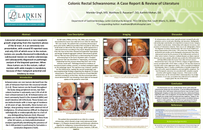
Case Description/Methods: A case of rectal schwannoma in an 85-year-old-female, who presented via referral for colonoscope evaluation of a previously identified lesion in the rectum concerning for malignancy. Pathology from prior deep biopsies revealed histopathology positive analysis for S-100 and negative for C-KIT, CD-34, Actin, and Desmin. Subsequent annual rectal EUS and FGD-PET scan surveillance was opted in by the patient instead of surgical removal. Follow-up for recurrence of lesion is ongoing with 6 to 12-month intervals.
Discussion: Gastrointestinal schwannomas occur indifferently in men and women with a mean age of 60–65 years. It can be incidentally diagnosed in an asymptomatic patient during a screening colonoscopy or with a wide range of symptomatic presentations, such as obstruction, bleeding, and tenesmus. Endoscopically, schwannomas tend to be lobulated well-defined tumors ulcerating into the mucosa. Pathologically, schwannomas are positive for S-100 and negative for CD117, Desmin, and actin. Histologically, there is a proliferation of spindle-shaped cells with oval nuclei and tapered ends, with eosinophilic cytoplasm with fuzzy boundaries. They are characterized by a low rate of mitosis, the absence of atypical mitotic figures, and nuclear hyperpigmentation. The degree of aggressiveness has been correlated with the Ki-67 index and the mitotic indices. Due to its risk of malignant transformation, resection of schwannoma is the recommended treatment. Clinical follow-up becomes very crucial. Little data exists on this rare entity in the literature with only a limited number of cases described. Highlighting these cases becomes paramount to help keep gastroenterologists more aware of this rare entity.
Disclosures:
Monider Singh, MD, MBA1, Matthew D.. Razavian, DO2, Aamir Pervez, DO1, Amir Riaz, MD1, Karthik Mohan, DO2. P2093 - Colonic Rectal Schwannoma: A Case Report and Review of Literature, ACG 2024 Annual Scientific Meeting Abstracts. Philadelphia, PA: American College of Gastroenterology.
1Larkin Community Hospital, South Miami, FL; 2Larkin Community Hospital, Hialeah, FL
Introduction: Colorectal schwannoma is a rare neoplastic growth, first described in 1962, originating from the myenteric plexus of the GI tract. The digestive and especially colonic location of this tumor is rare. More frequently in the stomach and the small intestine. It is an extremely rare presentation, with around 95 reported cases with only 21.1% of cases occurring in the rectum. The lesions are usually discovered incidentally, as submucosal masses on routine colonoscopy and subsequently diagnosed on pathologic examination of the operative specimen via immunohistochemical panel (S-100). When these tumors are in the colon and in the rectum, radical excision with wide margins is mandatory, due to their tendency to recur locally or become malignant, if left untreated.
Case Description/Methods: A case of rectal schwannoma in an 85-year-old-female, who presented via referral for colonoscope evaluation of a previously identified lesion in the rectum concerning for malignancy. Pathology from prior deep biopsies revealed histopathology positive analysis for S-100 and negative for C-KIT, CD-34, Actin, and Desmin. Subsequent annual rectal EUS and FGD-PET scan surveillance was opted in by the patient instead of surgical removal. Follow-up for recurrence of lesion is ongoing with 6 to 12-month intervals.
Discussion: Gastrointestinal schwannomas occur indifferently in men and women with a mean age of 60–65 years. It can be incidentally diagnosed in an asymptomatic patient during a screening colonoscopy or with a wide range of symptomatic presentations, such as obstruction, bleeding, and tenesmus. Endoscopically, schwannomas tend to be lobulated well-defined tumors ulcerating into the mucosa. Pathologically, schwannomas are positive for S-100 and negative for CD117, Desmin, and actin. Histologically, there is a proliferation of spindle-shaped cells with oval nuclei and tapered ends, with eosinophilic cytoplasm with fuzzy boundaries. They are characterized by a low rate of mitosis, the absence of atypical mitotic figures, and nuclear hyperpigmentation. The degree of aggressiveness has been correlated with the Ki-67 index and the mitotic indices. Due to its risk of malignant transformation, resection of schwannoma is the recommended treatment. Clinical follow-up becomes very crucial. Little data exists on this rare entity in the literature with only a limited number of cases described. Highlighting these cases becomes paramount to help keep gastroenterologists more aware of this rare entity.
Disclosures:
Monider Singh indicated no relevant financial relationships.
Matthew Razavian indicated no relevant financial relationships.
Aamir Pervez indicated no relevant financial relationships.
Amir Riaz indicated no relevant financial relationships.
Karthik Mohan indicated no relevant financial relationships.
Monider Singh, MD, MBA1, Matthew D.. Razavian, DO2, Aamir Pervez, DO1, Amir Riaz, MD1, Karthik Mohan, DO2. P2093 - Colonic Rectal Schwannoma: A Case Report and Review of Literature, ACG 2024 Annual Scientific Meeting Abstracts. Philadelphia, PA: American College of Gastroenterology.
