Monday Poster Session
Category: Esophagus
P2252 - Esophageal Stricture Post Endoscopic Submucosal Dissection of Distal Circumferential Esophageal Squamous Cell Dysplasia: A Persistent Challenge
Monday, October 28, 2024
10:30 AM - 4:00 PM ET
Location: Exhibit Hall E

Has Audio

Mohamad-Noor Abu-Hammour, MD
Cleveland Clinic Fairview
Cleveland, OH
Presenting Author(s)
Mohamad-Noor Abu-Hammour, MD1, Yi Qin, MD2, Siva Raja, MD3, Amit Bhatt, MD2
1Cleveland Clinic Fairview, Cleveland, OH; 2Digestive Disease Institute, Cleveland Clinic, Cleveland, OH; 3Heart and Vascular Institute, Cleveland Clinic, Cleveland, OH
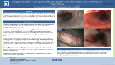
Introduction: Endoscopic submucosal dissection (ESD) has emerged as the preferred method for the treatment of superficial esophageal tumors. A challenge of ESD resection of esophageal tumors is when a large percentage of the lumen circumference is resected, there is a risk of esophageal stricture formation. We present a case of patient with a circumferential distal esophageal squamous cell dysplasia.
Case Description/Methods: A 68-year-old female with odynophagia. An upper endoscopy revealed a 5 cm circumferential distal esophageal tumor. Biopsies from the tumor revealed squamous cell carcinoma in situ. The patient was subsequently referred to our center for ESD.
Examination under both white light and narrow-band imaging (NBI) revealed approximately 5 cm of circumferential distal esophageal nodular mucosa with ulceration. Magnified NBI revealed intrapapillary capillary loops V1 and V2 suggesting superficial SCC. We theorized the ulceration may have been from reflux, rather than signifying a more advanced tumor, so proceeded with staging endoscopic submucosal dissection (ESD).
We used endoscopic tunneling submucosal dissection with IT tunneling technique. A mucosal incision was made at the proximal and distal end of the lesion using a needle-tip ESD knife. Then two submucosal tunnels extending from the proximal to distal mucosal incisions were created using IT tunneling knife technique. Clip line was then used for traction, and the remaining submucosal was dissected using an IT knife and the tumor was removed en-bloc. Purastat was applied to the resection base in hopes of reducing the risk of stricture formation.
Pathology following the procedure confirmed high-grade squamous dysplasia with negative margins. To prevent refractory stricture formation, the patient was advised to take twice daily proton pump inhibitor and a swallowed topical steroid daily.
Despite these preventive measures, the patient developed significant esophageal stenosis, necessitating multiple endoscopic balloon dilations (EBD).
Discussion: This case highlights the challenges of performing large esophageal ESD. Circumferential ESD resections, invariably result in esophageal stenosis despite use of multiple post-ESD stricture prevention methods. These post ESD stenoses can be recalcitrant, requiring multiple EBD sessions. Further research is required to develop novel methods for post ESD stricture prevention.

Disclosures:
Mohamad-Noor Abu-Hammour, MD1, Yi Qin, MD2, Siva Raja, MD3, Amit Bhatt, MD2. P2252 - Esophageal Stricture Post Endoscopic Submucosal Dissection of Distal Circumferential Esophageal Squamous Cell Dysplasia: A Persistent Challenge, ACG 2024 Annual Scientific Meeting Abstracts. Philadelphia, PA: American College of Gastroenterology.
1Cleveland Clinic Fairview, Cleveland, OH; 2Digestive Disease Institute, Cleveland Clinic, Cleveland, OH; 3Heart and Vascular Institute, Cleveland Clinic, Cleveland, OH
Introduction: Endoscopic submucosal dissection (ESD) has emerged as the preferred method for the treatment of superficial esophageal tumors. A challenge of ESD resection of esophageal tumors is when a large percentage of the lumen circumference is resected, there is a risk of esophageal stricture formation. We present a case of patient with a circumferential distal esophageal squamous cell dysplasia.
Case Description/Methods: A 68-year-old female with odynophagia. An upper endoscopy revealed a 5 cm circumferential distal esophageal tumor. Biopsies from the tumor revealed squamous cell carcinoma in situ. The patient was subsequently referred to our center for ESD.
Examination under both white light and narrow-band imaging (NBI) revealed approximately 5 cm of circumferential distal esophageal nodular mucosa with ulceration. Magnified NBI revealed intrapapillary capillary loops V1 and V2 suggesting superficial SCC. We theorized the ulceration may have been from reflux, rather than signifying a more advanced tumor, so proceeded with staging endoscopic submucosal dissection (ESD).
We used endoscopic tunneling submucosal dissection with IT tunneling technique. A mucosal incision was made at the proximal and distal end of the lesion using a needle-tip ESD knife. Then two submucosal tunnels extending from the proximal to distal mucosal incisions were created using IT tunneling knife technique. Clip line was then used for traction, and the remaining submucosal was dissected using an IT knife and the tumor was removed en-bloc. Purastat was applied to the resection base in hopes of reducing the risk of stricture formation.
Pathology following the procedure confirmed high-grade squamous dysplasia with negative margins. To prevent refractory stricture formation, the patient was advised to take twice daily proton pump inhibitor and a swallowed topical steroid daily.
Despite these preventive measures, the patient developed significant esophageal stenosis, necessitating multiple endoscopic balloon dilations (EBD).
Discussion: This case highlights the challenges of performing large esophageal ESD. Circumferential ESD resections, invariably result in esophageal stenosis despite use of multiple post-ESD stricture prevention methods. These post ESD stenoses can be recalcitrant, requiring multiple EBD sessions. Further research is required to develop novel methods for post ESD stricture prevention.

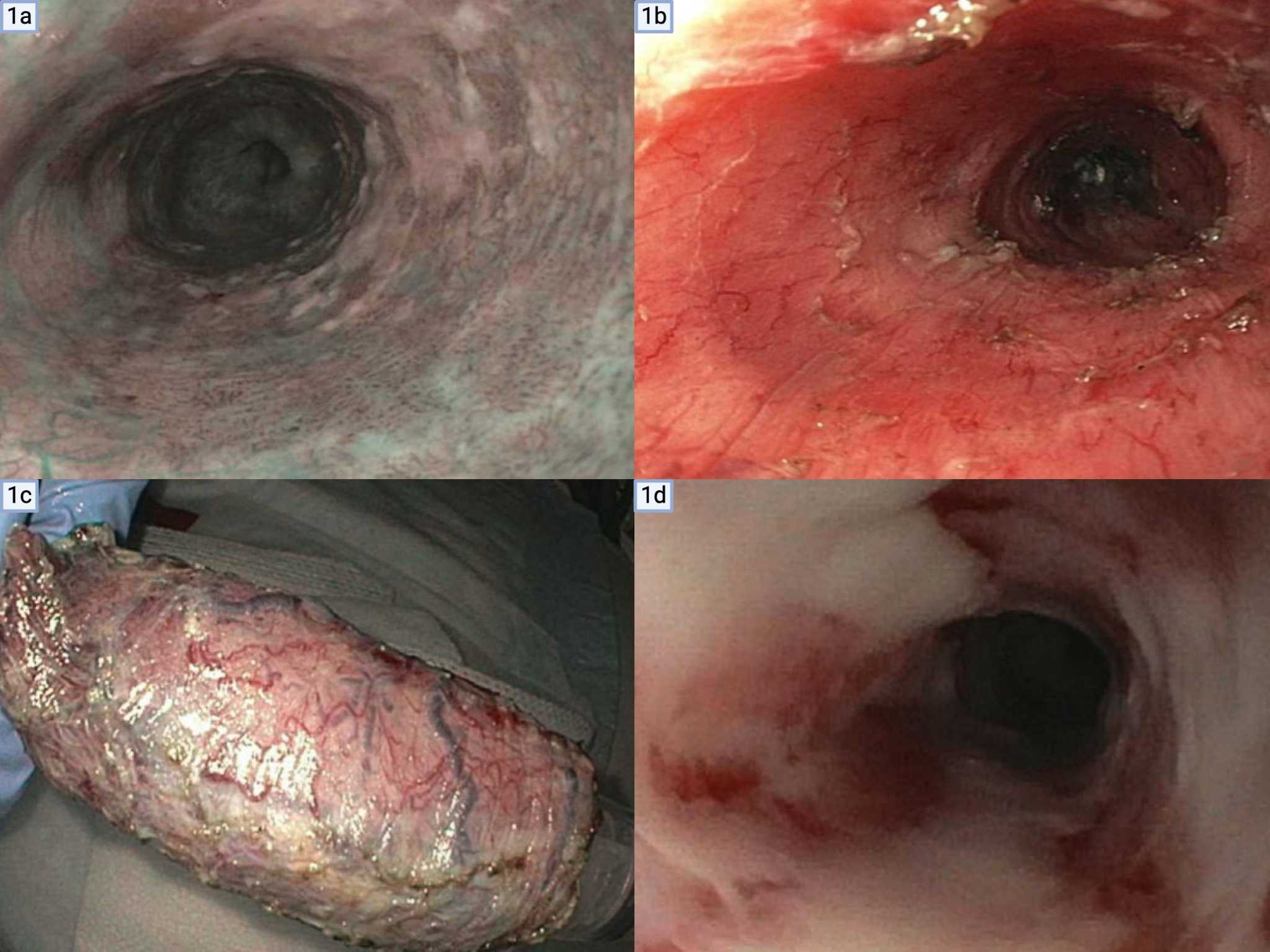
Figure: Figure 1 -
1a: Esophageal superficial tumor with ulceration
1b: Post-ESD resection in the esophagus
1c: Resected ESD specimen
1d: Post-ESD stricture
1a: Esophageal superficial tumor with ulceration
1b: Post-ESD resection in the esophagus
1c: Resected ESD specimen
1d: Post-ESD stricture
Disclosures:
Mohamad-Noor Abu-Hammour indicated no relevant financial relationships.
Yi Qin indicated no relevant financial relationships.
Siva Raja indicated no relevant financial relationships.
Amit Bhatt indicated no relevant financial relationships.
Mohamad-Noor Abu-Hammour, MD1, Yi Qin, MD2, Siva Raja, MD3, Amit Bhatt, MD2. P2252 - Esophageal Stricture Post Endoscopic Submucosal Dissection of Distal Circumferential Esophageal Squamous Cell Dysplasia: A Persistent Challenge, ACG 2024 Annual Scientific Meeting Abstracts. Philadelphia, PA: American College of Gastroenterology.
