Monday Poster Session
Category: Esophagus
P2253 - Uncovering the Unexpected: A Rare Case of Carcinoma Cuniculatum in the Esophagus
Monday, October 28, 2024
10:30 AM - 4:00 PM ET
Location: Exhibit Hall E

Has Audio

Lawrence Zhou, MD
The University of Kansas School of Medicine
Wichita, KS
Presenting Author(s)
Lawrence Zhou, MD1, Aastha V. Bharwad, MD2, Daly Al-Hadeethi, MD2, Kyle Rowe, MD3
1The University of Kansas School of Medicine, Wichita, KS; 2University of Kansas School of Medicine, Wichita, KS; 3University of Kansas Medical Center, Wichita, KS
Introduction: Carcinoma cuniculatum (CC) is a very rare variant of well-differentiated squamous cell carcinoma (SCC) of the oral cavity that is frequently misdiagnosed. Previous literature notes slight male predominance and risk factors similar to SCC, such as tobacco exposure via smoking or chewing, alcohol, betel leaf chewing, chronic reflux esophagitis, and immunosuppression. It is most common at the mandibular gingiva and often locally invasive, with a pathognomonic finding of bony invasion. Here we describe a middle-aged male found to have CC located in the upper thoracic esophagus.
Case Description/Methods: A 69 year-old male with a history of H. pylori gastritis and former smoking presents for three weeks difficulty swallowing solids progressing to liquids and unintentional weight loss.
Endogastroduodenoscopy (EGD) for dysphagia performed four years prior had revealed esophageal narrowing and inflammation at 20 cm requiring dilation and H. pylori gastritis. Biopsies did not show evidence of dysplasia or cancer.
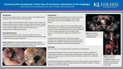
Repeat EGD showed a malignant appearing esophageal stricture spanning 20 to 25 cm arising in the background of severe squamous hyperplasia or epidermoid metaplasia (Figure A and B). Biopsies were consistent with CC.
Computed Tomography and positron emission tomography showed a primary esophageal neoplasm most prominent proximally and multiple hypermetabolic lymph nodes (Figure C and D). Bronchoscopy did not show endobronchial invasion. Carcinoembryonic antigen was elevated at 2.1 ng/mL.
Unfortunately, the patient was not deemed a surgical candidate, and oncology planned for chemoradiation with paclitaxel and carboplatin. Ultimately, the patient opted for hospice before passing two months after presentation.
Discussion: CC is easily missed or misdiagnosed due to its under-recognition as a variant of oral SCC, thus leading to risk for delayed treatment. Characteristic histologic attributes include hyperkeratosis, acanthosis, dyskeratosis, intraepithelial neutrophils, microabscesses, cytologic atypia, deep keratinization, and koilocytic-like squamous cells. These attributes have been summarized as resembling ‘rabbit burrows’. If identified early, definitive radiotherapy or primary resection alone may be enough, or in this case of locoregionally advanced disease treated with multiple modalities of surgery, radiotherapy, and/or chemotherapy.

Disclosures:
Lawrence Zhou, MD1, Aastha V. Bharwad, MD2, Daly Al-Hadeethi, MD2, Kyle Rowe, MD3. P2253 - Uncovering the Unexpected: A Rare Case of Carcinoma Cuniculatum in the Esophagus, ACG 2024 Annual Scientific Meeting Abstracts. Philadelphia, PA: American College of Gastroenterology.
1The University of Kansas School of Medicine, Wichita, KS; 2University of Kansas School of Medicine, Wichita, KS; 3University of Kansas Medical Center, Wichita, KS
Introduction: Carcinoma cuniculatum (CC) is a very rare variant of well-differentiated squamous cell carcinoma (SCC) of the oral cavity that is frequently misdiagnosed. Previous literature notes slight male predominance and risk factors similar to SCC, such as tobacco exposure via smoking or chewing, alcohol, betel leaf chewing, chronic reflux esophagitis, and immunosuppression. It is most common at the mandibular gingiva and often locally invasive, with a pathognomonic finding of bony invasion. Here we describe a middle-aged male found to have CC located in the upper thoracic esophagus.
Case Description/Methods: A 69 year-old male with a history of H. pylori gastritis and former smoking presents for three weeks difficulty swallowing solids progressing to liquids and unintentional weight loss.
Endogastroduodenoscopy (EGD) for dysphagia performed four years prior had revealed esophageal narrowing and inflammation at 20 cm requiring dilation and H. pylori gastritis. Biopsies did not show evidence of dysplasia or cancer.
Repeat EGD showed a malignant appearing esophageal stricture spanning 20 to 25 cm arising in the background of severe squamous hyperplasia or epidermoid metaplasia (Figure A and B). Biopsies were consistent with CC.
Computed Tomography and positron emission tomography showed a primary esophageal neoplasm most prominent proximally and multiple hypermetabolic lymph nodes (Figure C and D). Bronchoscopy did not show endobronchial invasion. Carcinoembryonic antigen was elevated at 2.1 ng/mL.
Unfortunately, the patient was not deemed a surgical candidate, and oncology planned for chemoradiation with paclitaxel and carboplatin. Ultimately, the patient opted for hospice before passing two months after presentation.
Discussion: CC is easily missed or misdiagnosed due to its under-recognition as a variant of oral SCC, thus leading to risk for delayed treatment. Characteristic histologic attributes include hyperkeratosis, acanthosis, dyskeratosis, intraepithelial neutrophils, microabscesses, cytologic atypia, deep keratinization, and koilocytic-like squamous cells. These attributes have been summarized as resembling ‘rabbit burrows’. If identified early, definitive radiotherapy or primary resection alone may be enough, or in this case of locoregionally advanced disease treated with multiple modalities of surgery, radiotherapy, and/or chemotherapy.

Figure: Figure A - Upper third of the esophagus, view #1. Figure B - Upper third of the esophagus, view #2. Figure C - Positron emission tomography, coronal view. Figure D - Positron emission tomography, axial view.
Disclosures:
Lawrence Zhou indicated no relevant financial relationships.
Aastha Bharwad indicated no relevant financial relationships.
Daly Al-Hadeethi indicated no relevant financial relationships.
Kyle Rowe indicated no relevant financial relationships.
Lawrence Zhou, MD1, Aastha V. Bharwad, MD2, Daly Al-Hadeethi, MD2, Kyle Rowe, MD3. P2253 - Uncovering the Unexpected: A Rare Case of Carcinoma Cuniculatum in the Esophagus, ACG 2024 Annual Scientific Meeting Abstracts. Philadelphia, PA: American College of Gastroenterology.
