Monday Poster Session
Category: Esophagus
P2256 - Chronic Aspiration: A Potential Complication of Cricopharyngeal Muscle Dysfunction (CMD)
Monday, October 28, 2024
10:30 AM - 4:00 PM ET
Location: Exhibit Hall E

Has Audio
- AK
Andrew Kim, MD
Walter Reed National Military Medical Center
Bethesda, MD
Presenting Author(s)
Andrew Kim, MD, William F. Kelly, MD, Addison F. Jacobs, MD, Dean E. Baird, MD, Roy K. Wong, MD, MACG
Walter Reed National Military Medical Center, Bethesda, MD
Introduction: Cricopharyngeal muscle dysfunction (CMD) is a narrowing of the upper esophageal sphincter (UES) that occurs when there is incomplete relaxation of the muscle resulting in oropharyngeal dysphagia. It is thought that the partial replacement of cricopharyngeal (CP) muscle with fibrous connective tissue results in decreased CP muscle compliance and luminal diameter that may appear radiologically as a CP bar. We report a case of a 75 year old female with chronic cough / aspiration pneumonitis in setting of dysphagia / CMD that improves dramatically with large diameter esophageal dilations.
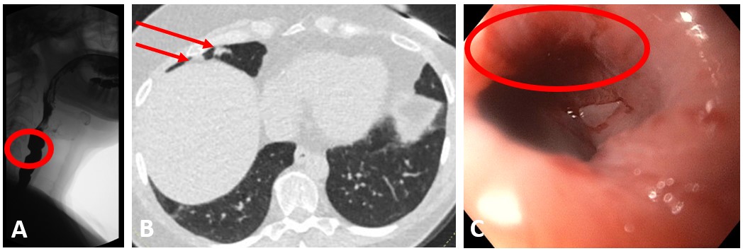
Case Description/Methods: 75 year old female complaining of solid-food dysphagia for over two decades in the setting of CP bar (figure A), normal EGD with esophageal biopsies, and normal esophageal motility studies. Prior to interval dilations, she complained of pills getting stuck in her upper throat with associated coughing / shortness of breath, endorsing behavior modification such as finely cutting solid food in her meals. Swallowing and coughing dramatically improved for up to 3 years with interval dilations greater than 50 French. Interval non-contrast chest CT demonstrated radiographic improvement of chronic changes suspected secondary to chronic aspiration (figure B). Small caliber esophageal dilations less than 50 French resulted in 1-2 weeks of symptom relief. Her most recent Maloney dilation with 60 French resulted in a CP tear with excellent relief of symptoms (figure C).
Discussion: Increased CP fibrosis with luminal narrowing is the probable cause of her oropharyngeal dysphagia as evidence by the mucosal tear of the CP muscle and subsequent long-term efficacy with large diameter dilations (greater than 50 French). The CP fibrotic tissue needs to be disrupted. This case highlights potential preventable chronic manifestations of CMD, specifically chronic aspiration. Finally, given that not all patients with CMD have a CP bar, the case suggests considering undiagnosed CMD in patients with oropharyngeal dysphagia and idiopathic chronic aspiration.

Disclosures:
Andrew Kim, MD, William F. Kelly, MD, Addison F. Jacobs, MD, Dean E. Baird, MD, Roy K. Wong, MD, MACG. P2256 - Chronic Aspiration: A Potential Complication of Cricopharyngeal Muscle Dysfunction (CMD), ACG 2024 Annual Scientific Meeting Abstracts. Philadelphia, PA: American College of Gastroenterology.
Walter Reed National Military Medical Center, Bethesda, MD
Introduction: Cricopharyngeal muscle dysfunction (CMD) is a narrowing of the upper esophageal sphincter (UES) that occurs when there is incomplete relaxation of the muscle resulting in oropharyngeal dysphagia. It is thought that the partial replacement of cricopharyngeal (CP) muscle with fibrous connective tissue results in decreased CP muscle compliance and luminal diameter that may appear radiologically as a CP bar. We report a case of a 75 year old female with chronic cough / aspiration pneumonitis in setting of dysphagia / CMD that improves dramatically with large diameter esophageal dilations.
Case Description/Methods: 75 year old female complaining of solid-food dysphagia for over two decades in the setting of CP bar (figure A), normal EGD with esophageal biopsies, and normal esophageal motility studies. Prior to interval dilations, she complained of pills getting stuck in her upper throat with associated coughing / shortness of breath, endorsing behavior modification such as finely cutting solid food in her meals. Swallowing and coughing dramatically improved for up to 3 years with interval dilations greater than 50 French. Interval non-contrast chest CT demonstrated radiographic improvement of chronic changes suspected secondary to chronic aspiration (figure B). Small caliber esophageal dilations less than 50 French resulted in 1-2 weeks of symptom relief. Her most recent Maloney dilation with 60 French resulted in a CP tear with excellent relief of symptoms (figure C).
Discussion: Increased CP fibrosis with luminal narrowing is the probable cause of her oropharyngeal dysphagia as evidence by the mucosal tear of the CP muscle and subsequent long-term efficacy with large diameter dilations (greater than 50 French). The CP fibrotic tissue needs to be disrupted. This case highlights potential preventable chronic manifestations of CMD, specifically chronic aspiration. Finally, given that not all patients with CMD have a CP bar, the case suggests considering undiagnosed CMD in patients with oropharyngeal dysphagia and idiopathic chronic aspiration.

Figure: Figure A is a barium swallow demonstrating a small cricopharyngeal bar (circled) at the level of C5. Figure B is a non-contrast chest CT demonstrating right middle lobe nodularity, PET avid, waxing and waning over time (arrows) that resolved (and developed elsewhere), suspected secondary to aspiration. Figure C demonstrates esophageal mucosal tearing (circled) after her most recent large caliber dilation.
Disclosures:
Andrew Kim indicated no relevant financial relationships.
William Kelly indicated no relevant financial relationships.
Addison Jacobs indicated no relevant financial relationships.
Dean Baird indicated no relevant financial relationships.
Roy Wong indicated no relevant financial relationships.
Andrew Kim, MD, William F. Kelly, MD, Addison F. Jacobs, MD, Dean E. Baird, MD, Roy K. Wong, MD, MACG. P2256 - Chronic Aspiration: A Potential Complication of Cricopharyngeal Muscle Dysfunction (CMD), ACG 2024 Annual Scientific Meeting Abstracts. Philadelphia, PA: American College of Gastroenterology.

