Monday Poster Session
Category: Esophagus
P2270 - A Rare Case of Dual Pathogen Infectious Esophagitis
Monday, October 28, 2024
10:30 AM - 4:00 PM ET
Location: Exhibit Hall E

Has Audio
- CP
Chandler Patton, DO
Lehigh Valley Health Network
Allentown, PA
Presenting Author(s)
Chandler Patton, DO1, Corey Savard, MD2, Timothy Graziano, DO1, Amanda Jacubowsky, DO1, Shashin Shah, MD3
1Lehigh Valley Health Network, Allentown, PA; 2Lehigh Valley Health Network -, Allentown, PA; 3Eastern Pennsylvania Gastrointestinal and Liver Specialists, Allentown, PA
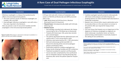
Introduction: Infectious esophagitis is a disease that predominantly affects immunocompromised patients. The most common causes of infectious esophagitis are Candida, HSV, and CMV. Bacterial pathogens are an uncommon cause of infectious esophagitis. It is rare for infectious esophagitis to be caused by two pathogens simultaneously with only a few cases in literature reporting this phenomenon. Here we present a case of concurrent Candida and HSV esophagitis in an immunocompromised patient undergoing chemotherapy and high dose steroids.
Case Description/Methods: A 39 year old male with a history of metastatic colon cancer on FOLFOX and Nivolumab and GERD presented with a full body blistering rash. On arrival, the patient was hemodynamically stable with labs significant for pancytopenia with neutropenia (absolute neutrophil count of 0.5 thou/cmm) and leukopenia (0.8 thou/cmm). The patient was evaluated by the dermatology team and skin biopsy performed. He was diagnosed with SJS vs TEN secondary to Nivolumab. He was treated with Etanercept, IVIG, pulse dose steroids with steroid taper. Two weeks later his hospital course was complicated by abdominal pain and melena and CT imaging was performed with findings concerning for colitis. Infectious stool panel showed C.Diff colonization. EGD and colonoscopy were performed (absolute neutrophil count of 3.4 thou/cmm) and exam revealed white oral and esophageal plaques concerning for candida esophagitis and biopsy was performed. Patient was started on empiric treatment for presumed candida esophagitis. Biopsy stains were significant for concomitant Candida and HSV. He was treated with Acyclovir in addition to Fluconazole. Their hospital course was complicated by microperforation from extensive metastatic disease requiring a diverting loop ileostomy and the decision was made to pursue hospice.
Discussion: This case demonstrates a patient with multiple mechanisms for immunosuppression found to have simultaneous Candida and HSV esophagitis. Candida esophagitis presents with characteristic white plaques on endoscopic exams often leading to empiric treatment without biopsy confirmed diagnosis. Biopsy is often avoided in these high risk patients due to factors such as neutropenia, and thrombocytopenia. This case demonstrates the importance of biopsy for definitive diagnosis and to rule out coinfection. Without biopsy this patient may have required repeat interventions and delay in proper treatment.

Disclosures:
Chandler Patton, DO1, Corey Savard, MD2, Timothy Graziano, DO1, Amanda Jacubowsky, DO1, Shashin Shah, MD3. P2270 - A Rare Case of Dual Pathogen Infectious Esophagitis, ACG 2024 Annual Scientific Meeting Abstracts. Philadelphia, PA: American College of Gastroenterology.
1Lehigh Valley Health Network, Allentown, PA; 2Lehigh Valley Health Network -, Allentown, PA; 3Eastern Pennsylvania Gastrointestinal and Liver Specialists, Allentown, PA
Introduction: Infectious esophagitis is a disease that predominantly affects immunocompromised patients. The most common causes of infectious esophagitis are Candida, HSV, and CMV. Bacterial pathogens are an uncommon cause of infectious esophagitis. It is rare for infectious esophagitis to be caused by two pathogens simultaneously with only a few cases in literature reporting this phenomenon. Here we present a case of concurrent Candida and HSV esophagitis in an immunocompromised patient undergoing chemotherapy and high dose steroids.
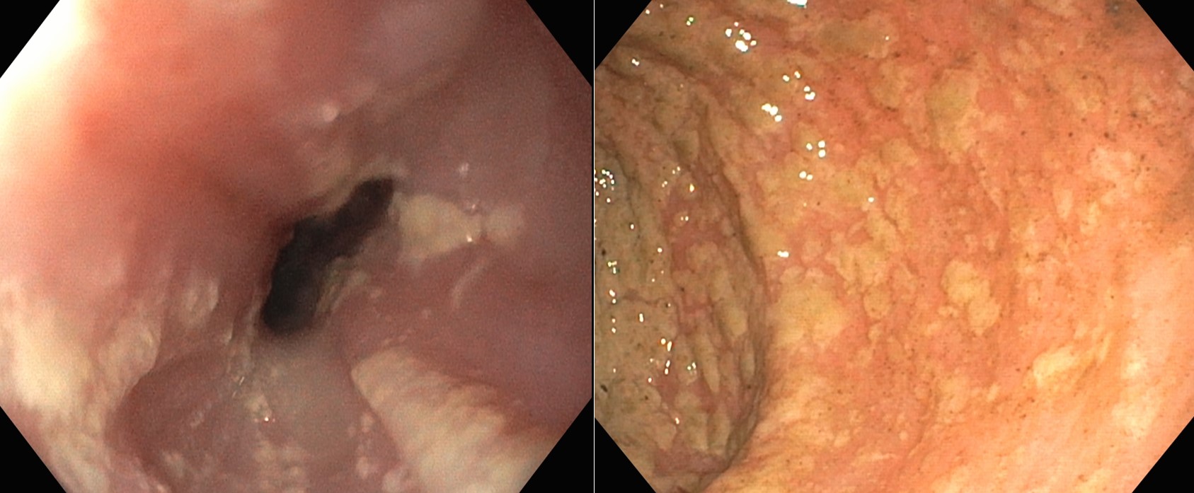
Case Description/Methods: A 39 year old male with a history of metastatic colon cancer on FOLFOX and Nivolumab and GERD presented with a full body blistering rash. On arrival, the patient was hemodynamically stable with labs significant for pancytopenia with neutropenia (absolute neutrophil count of 0.5 thou/cmm) and leukopenia (0.8 thou/cmm). The patient was evaluated by the dermatology team and skin biopsy performed. He was diagnosed with SJS vs TEN secondary to Nivolumab. He was treated with Etanercept, IVIG, pulse dose steroids with steroid taper. Two weeks later his hospital course was complicated by abdominal pain and melena and CT imaging was performed with findings concerning for colitis. Infectious stool panel showed C.Diff colonization. EGD and colonoscopy were performed (absolute neutrophil count of 3.4 thou/cmm) and exam revealed white oral and esophageal plaques concerning for candida esophagitis and biopsy was performed. Patient was started on empiric treatment for presumed candida esophagitis. Biopsy stains were significant for concomitant Candida and HSV. He was treated with Acyclovir in addition to Fluconazole. Their hospital course was complicated by microperforation from extensive metastatic disease requiring a diverting loop ileostomy and the decision was made to pursue hospice.
Discussion: This case demonstrates a patient with multiple mechanisms for immunosuppression found to have simultaneous Candida and HSV esophagitis. Candida esophagitis presents with characteristic white plaques on endoscopic exams often leading to empiric treatment without biopsy confirmed diagnosis. Biopsy is often avoided in these high risk patients due to factors such as neutropenia, and thrombocytopenia. This case demonstrates the importance of biopsy for definitive diagnosis and to rule out coinfection. Without biopsy this patient may have required repeat interventions and delay in proper treatment.

Figure: Figure A and B: White plaques and exudates throughout the esophagus
Disclosures:
Chandler Patton indicated no relevant financial relationships.
Corey Savard indicated no relevant financial relationships.
Timothy Graziano indicated no relevant financial relationships.
Amanda Jacubowsky indicated no relevant financial relationships.
Shashin Shah indicated no relevant financial relationships.
Chandler Patton, DO1, Corey Savard, MD2, Timothy Graziano, DO1, Amanda Jacubowsky, DO1, Shashin Shah, MD3. P2270 - A Rare Case of Dual Pathogen Infectious Esophagitis, ACG 2024 Annual Scientific Meeting Abstracts. Philadelphia, PA: American College of Gastroenterology.
