Monday Poster Session
Category: Esophagus
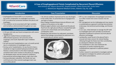
P2272 - A Case of Esophagopleural Fistula Complicated by Recurrent Pleural Effusions
Monday, October 28, 2024
10:30 AM - 4:00 PM ET
Location: Exhibit Hall E

Has Audio

Mark DeSantis, DO, MS
AtlantiCare Regional Medical Center
Brigantine, NJ
Presenting Author(s)
Mark DeSantis, DO, MS, Mamun Ahmed, DO, Deepika Davalath, MD, Nikita Chintam, MD, Sunita Yadav, MD
AtlantiCare Regional Medical Center, Atlantic City, NJ
Introduction: Esophagopleural fistula (EPF) is an uncommon but severe complication of esophageal carcinoma. EPF is an abnormal connection between the pleura and esophagus. This condition can be associated with high morbidity and mortality as demonstrated throughout our case.
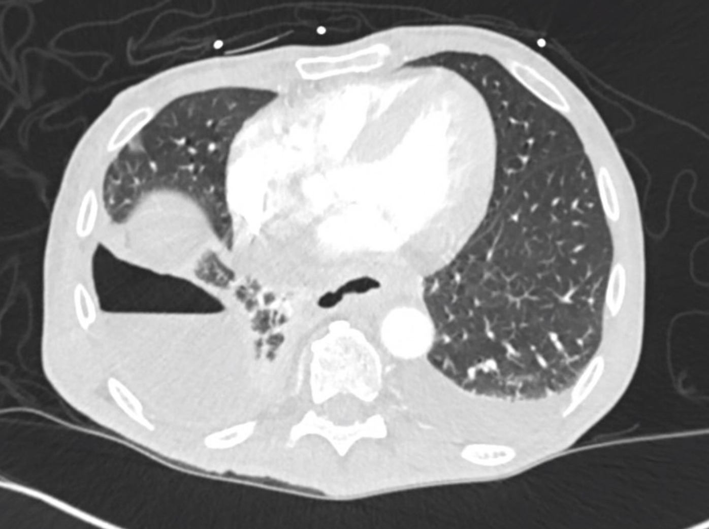
Case Description/Methods: A 59-year-old male was found unresponsive with severe hypoglycemia. Significant medical history included poorly differentiated squamous cell carcinoma of the esophagus and insulin dependent type II diabetes mellitus. He had recurrent hospitalizations over a 6-month period due to complications of esophageal cancer. During a prior hospitalization, he was found to have a moderate to large loculated hydropneumothorax with fistulous communication to the distal esophagus consistent with tumor invasion to the pleura on imaging. He developed sepsis and was started on antibiotics. Interventional pulmonology placed a right chest tube. During endoscopic evaluation, the fistula tract was confirmed and a fully covered esophageal stent was successfully placed. His clinical condition improved and he was discharged. A few weeks later, he presented due to hypoglycemia and signs of sepsis. New imaging revealed bilateral pleural effusions and evidence of loculation on the right. Bilateral pigtail catheters were placed and the pleural fluid from the right pleural effusion revealed fluid with concerns for enteral feeds and eventually grew Lactobacillus caseii. Despite the interventions he underwent, persistent pleural fluid leakage necessitated the placement of a permanent pleural catheter system.
Discussion: EPF is a rare complication of esophageal carcinoma as it often results from tumor invasion into the pleura. EPF management can be challenging and may require a multidisciplinary approach involving interventional endoscopy, interventional pulmonology, and thoracic surgery. In our case, initial attempts to manage the EPF with an esophageal stent were partially successful but ultimately insufficient to prevent recurrent pleural effusions and leakage. Lactobacillus caseii found in the pleural fluid is unusual and raises concern for enteral feeding refluxing through the fistula tract then contributing to subsequent infection and sepsis. The complexities in this case emphasize the importance of careful monitoring and a multidisciplinary approach to optimize patient outcomes. Further research is warranted to investigate optimal management strategies for patients who present with similar presentations.

Disclosures:
Mark DeSantis, DO, MS, Mamun Ahmed, DO, Deepika Davalath, MD, Nikita Chintam, MD, Sunita Yadav, MD. P2272 - A Case of Esophagopleural Fistula Complicated by Recurrent Pleural Effusions, ACG 2024 Annual Scientific Meeting Abstracts. Philadelphia, PA: American College of Gastroenterology.
AtlantiCare Regional Medical Center, Atlantic City, NJ
Introduction: Esophagopleural fistula (EPF) is an uncommon but severe complication of esophageal carcinoma. EPF is an abnormal connection between the pleura and esophagus. This condition can be associated with high morbidity and mortality as demonstrated throughout our case.
Case Description/Methods: A 59-year-old male was found unresponsive with severe hypoglycemia. Significant medical history included poorly differentiated squamous cell carcinoma of the esophagus and insulin dependent type II diabetes mellitus. He had recurrent hospitalizations over a 6-month period due to complications of esophageal cancer. During a prior hospitalization, he was found to have a moderate to large loculated hydropneumothorax with fistulous communication to the distal esophagus consistent with tumor invasion to the pleura on imaging. He developed sepsis and was started on antibiotics. Interventional pulmonology placed a right chest tube. During endoscopic evaluation, the fistula tract was confirmed and a fully covered esophageal stent was successfully placed. His clinical condition improved and he was discharged. A few weeks later, he presented due to hypoglycemia and signs of sepsis. New imaging revealed bilateral pleural effusions and evidence of loculation on the right. Bilateral pigtail catheters were placed and the pleural fluid from the right pleural effusion revealed fluid with concerns for enteral feeds and eventually grew Lactobacillus caseii. Despite the interventions he underwent, persistent pleural fluid leakage necessitated the placement of a permanent pleural catheter system.
Discussion: EPF is a rare complication of esophageal carcinoma as it often results from tumor invasion into the pleura. EPF management can be challenging and may require a multidisciplinary approach involving interventional endoscopy, interventional pulmonology, and thoracic surgery. In our case, initial attempts to manage the EPF with an esophageal stent were partially successful but ultimately insufficient to prevent recurrent pleural effusions and leakage. Lactobacillus caseii found in the pleural fluid is unusual and raises concern for enteral feeding refluxing through the fistula tract then contributing to subsequent infection and sepsis. The complexities in this case emphasize the importance of careful monitoring and a multidisciplinary approach to optimize patient outcomes. Further research is warranted to investigate optimal management strategies for patients who present with similar presentations.

Figure: Image 1: CTA Chest showing loculated hydropneumothorax with fistulous communication between distal esophagus and pleura
Disclosures:
Mark DeSantis indicated no relevant financial relationships.
Mamun Ahmed indicated no relevant financial relationships.
Deepika Davalath indicated no relevant financial relationships.
Nikita Chintam indicated no relevant financial relationships.
Sunita Yadav indicated no relevant financial relationships.
Mark DeSantis, DO, MS, Mamun Ahmed, DO, Deepika Davalath, MD, Nikita Chintam, MD, Sunita Yadav, MD. P2272 - A Case of Esophagopleural Fistula Complicated by Recurrent Pleural Effusions, ACG 2024 Annual Scientific Meeting Abstracts. Philadelphia, PA: American College of Gastroenterology.
