Monday Poster Session
Category: Esophagus
P2291 - Herpes Simplex Virus and Cytomegalovirus Coinfection of the Esophagus in an Immunosuppressed Patient
Monday, October 28, 2024
10:30 AM - 4:00 PM ET
Location: Exhibit Hall E

Has Audio
- RB
Rishi Bolla, MD
University of South Florida Morsani College of Medicine
Tampa, FL
Presenting Author(s)
Rishi Bolla, MD1, Liza Plafsky, MD2, David Metter, MD3, John Jacobs, MD, FACG1
1University of South Florida Morsani College of Medicine, Tampa, FL; 2University of South Florida, Tampa, FL; 3Tampa General Hospital / University of South Florida, Tampa, FL
Introduction: Herpes simplex virus (HSV) and cytomegalovirus (CMV) are common causes of viral esophagitis. It is a well-known complication in patients with organ transplantation as immunosuppression is a major risk factor. Here we present a rare case of HSV/CMV coinfection which developed rapidly and caused severe esophagitis in an immunosuppressed patient.
Case Description/Methods: A 71-year-old male with past medical history of IgA nephropathy status post kidney transplant five years ago – on tacrolimus, mycophenolate mofetil, and daily prednisone – was evaluated for iron deficiency anemia. Six months prior, the patient was anemic in the setting of active melena, and he underwent esophagogastroduodenoscopy (EGD) and was treated for acute gastritis with active hemorrhage and a Forrest Class III duodenal ulcer. The esophagus was normal. Colonoscopy was attempted but aborted due to poor bowel preparation.
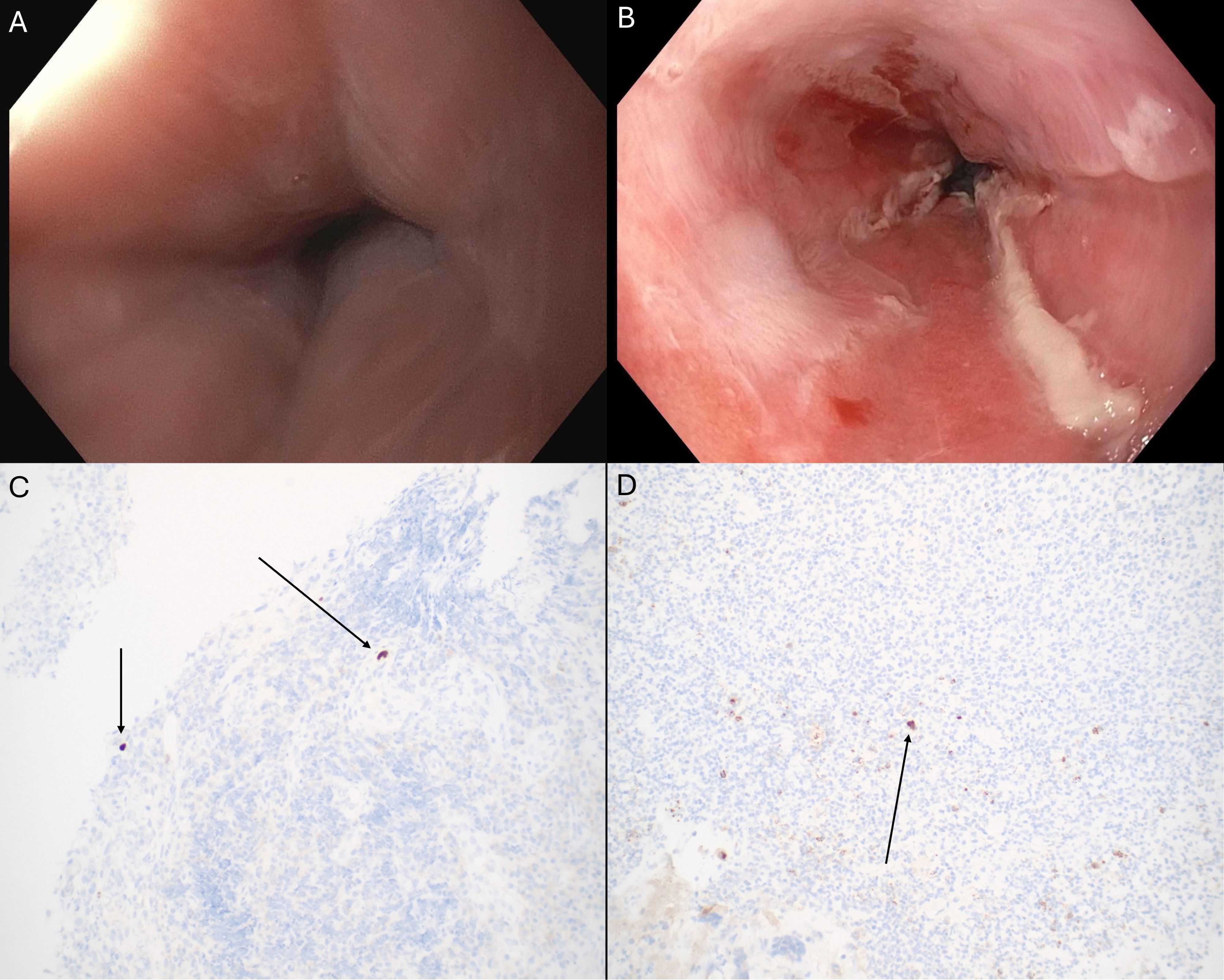
Since then, the patient remained anemic without active bleeding. He complained of intermittent bloating and abdominal pain but denied any dysphagia/odynophagia. Repeat EGD and colonoscopy were planned. Colonoscopy showed no abnormal findings, albeit with poor preparation once again. EGD however revealed severe LA Grade D esophagitis in the distal esophagus with ulceration and overlying exudate. Biopsies were positive for both HSV and CMV by immunohistochemistry. He was initiated on a valganciclovir course for six weeks and instructed to follow up with infectious disease and transplant nephrology specialists.
Discussion: HSV and CMV coinfection of the esophagus is a rare phenomenon. To our knowledge, there are few cases of HSV/CMV co-infection esophagitis in the literature (1,2). Notably, our patient did not complain of any esophageal symptoms such as dysphagia/odynophagia or reflux. Additionally, the patient’s clinical course illustrates how rapidly HSV/CMV esophagitis can develop. EGD merely six months before revealed a normal esophagus, but by the time of the current presentation, he had developed severe HSV/CMV esophagitis.
References
1. Bannoura et al. Esophageal Cytomegalovirus and Herpes Simplex virus co-infection in an immunocompromised patient: Case report and review of literature. IDCases. 2020 Jan 1;22:e00925.
2. Gilbert WC, Shah A. S2130 A Hard Case to Swallow: Co-Infectious Viral Esophagitis as a Complication of Steroid Use for Alcoholic Hepatitis. Official journal of the American College of Gastroenterology| ACG. 2021 Oct 1;116:S916.

Disclosures:
Rishi Bolla, MD1, Liza Plafsky, MD2, David Metter, MD3, John Jacobs, MD, FACG1. P2291 - Herpes Simplex Virus and Cytomegalovirus Coinfection of the Esophagus in an Immunosuppressed Patient, ACG 2024 Annual Scientific Meeting Abstracts. Philadelphia, PA: American College of Gastroenterology.
1University of South Florida Morsani College of Medicine, Tampa, FL; 2University of South Florida, Tampa, FL; 3Tampa General Hospital / University of South Florida, Tampa, FL
Introduction: Herpes simplex virus (HSV) and cytomegalovirus (CMV) are common causes of viral esophagitis. It is a well-known complication in patients with organ transplantation as immunosuppression is a major risk factor. Here we present a rare case of HSV/CMV coinfection which developed rapidly and caused severe esophagitis in an immunosuppressed patient.
Case Description/Methods: A 71-year-old male with past medical history of IgA nephropathy status post kidney transplant five years ago – on tacrolimus, mycophenolate mofetil, and daily prednisone – was evaluated for iron deficiency anemia. Six months prior, the patient was anemic in the setting of active melena, and he underwent esophagogastroduodenoscopy (EGD) and was treated for acute gastritis with active hemorrhage and a Forrest Class III duodenal ulcer. The esophagus was normal. Colonoscopy was attempted but aborted due to poor bowel preparation.
Since then, the patient remained anemic without active bleeding. He complained of intermittent bloating and abdominal pain but denied any dysphagia/odynophagia. Repeat EGD and colonoscopy were planned. Colonoscopy showed no abnormal findings, albeit with poor preparation once again. EGD however revealed severe LA Grade D esophagitis in the distal esophagus with ulceration and overlying exudate. Biopsies were positive for both HSV and CMV by immunohistochemistry. He was initiated on a valganciclovir course for six weeks and instructed to follow up with infectious disease and transplant nephrology specialists.
Discussion: HSV and CMV coinfection of the esophagus is a rare phenomenon. To our knowledge, there are few cases of HSV/CMV co-infection esophagitis in the literature (1,2). Notably, our patient did not complain of any esophageal symptoms such as dysphagia/odynophagia or reflux. Additionally, the patient’s clinical course illustrates how rapidly HSV/CMV esophagitis can develop. EGD merely six months before revealed a normal esophagus, but by the time of the current presentation, he had developed severe HSV/CMV esophagitis.
References
1. Bannoura et al. Esophageal Cytomegalovirus and Herpes Simplex virus co-infection in an immunocompromised patient: Case report and review of literature. IDCases. 2020 Jan 1;22:e00925.
2. Gilbert WC, Shah A. S2130 A Hard Case to Swallow: Co-Infectious Viral Esophagitis as a Complication of Steroid Use for Alcoholic Hepatitis. Official journal of the American College of Gastroenterology| ACG. 2021 Oct 1;116:S916.

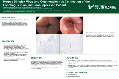
Figure: Figure 1: Distal esophagus visualized six months prior via EGD (A) versus at time of presentation (B). Immunohistochemistry stains at 20x magnification of the distal esophageal biopsies with brown nuclear staining in large cells positive for CMV (C) and HSV (D).
Disclosures:
Rishi Bolla indicated no relevant financial relationships.
Liza Plafsky indicated no relevant financial relationships.
David Metter indicated no relevant financial relationships.
John Jacobs: Medtronic – Consultant. Regeneron – Speakers Bureau. Sanofi/Genzyme – Speakers Bureau.
Rishi Bolla, MD1, Liza Plafsky, MD2, David Metter, MD3, John Jacobs, MD, FACG1. P2291 - Herpes Simplex Virus and Cytomegalovirus Coinfection of the Esophagus in an Immunosuppressed Patient, ACG 2024 Annual Scientific Meeting Abstracts. Philadelphia, PA: American College of Gastroenterology.
