Monday Poster Session
Category: General Endoscopy
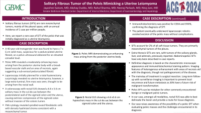
P2421 - Solitary Fibrous Tumor of the Pelvis Mimicking a Uterine Leiomyoma
Monday, October 28, 2024
10:30 AM - 4:00 PM ET
Location: Exhibit Hall E

Has Audio

Jayashrei Sairam, MD
Greater Baltimore Medical Center
Towson, MD
Presenting Author(s)
Jayashrei Sairam, MD, Akshay Duddu, MD, Rahul Sharma, MD, Neeraj Parkash, MD, Niraj Jani, MD
Greater Baltimore Medical Center, Towson, MD
Introduction: Solitary fibrous tumors (SFTs) are rare mesenchymal tumors, mainly of the pleural space, with an annual incidence of 1 case/million people. Here, we report a rare case of SFT of the pelvis that was initially diagnosed as uterine leiomyoma.
Case Description/Methods: A 40-year-old transgender man was found to have a 7.3x 6 cm solid mass, suspicious for a pedunculated uterine fibroid on a pelvic ultrasound obtained for evaluation of elevated estradiol level. Pelvic MRI revealed a moderately enhancing mass arising from the posterior uterine body with a broad-based vascular stalk and an area of central necrosis, suggestive of a subserosal pedunculated fibroid. Laparoscopy initially planned for hysterectomy surprisingly revealed no uterine leiomyoma; however, a large extra-luminal mass was seen, thought to be arising from the rectal wall. A colonoscopy with rectal EUS showed a 6 x 5.6 cm solitary mass in the cul-de-sac between the posterolateral wall of the sigmoid colon and the uterus, causing extrinsic compression of the sigmoid colon without invasion of the colonic lumen. FNA cytology revealed spindled-ovoid fibroblastic cells with densely hyalinized stroma consistent with a mesenchymal tumor with no cellular atypia or significant mitotic activity. Immunohistochemistry was positive for CD34 and STAT6, confirming the diagnosis of SFT. The patient underwent laparoscopic robotic-assisted excision of the pelvis mass without complications.
Discussion: SFTs account for 2% of all soft tissue tumors. They are primarily mesenchymal tumors of the pleura. Extra-thoracic SFTs are extremely rare with abdominopelvic SFTs only described in case reports. Definitive diagnosis is based on characteristic microscopic appearance and immunohistochemical staining pattern. On imaging, features of heterogenous enhancement with areas of necrosis aid with the diagnosis, though not pathognomonic of the disease. The mainstay of treatment is surgical resection. Long-term follow-up with surveillance imaging is important to prevent local recurrence and future metastasis as 20% of these tumors have a risk of malignant transformation. Pelvic SFTs can be mistaken for other commonly encountered benign or malignant pelvic tumors. In our case, amongst all modalities, rectal EUS was able to best delineate the lesion in addition to providing a route for tissue diagnosis. Our case raises awareness of the possibility of a pelvic SFT when evaluating pelvic masses and the challenges encountered in its diagnosis.

Disclosures:
Jayashrei Sairam, MD, Akshay Duddu, MD, Rahul Sharma, MD, Neeraj Parkash, MD, Niraj Jani, MD. P2421 - Solitary Fibrous Tumor of the Pelvis Mimicking a Uterine Leiomyoma, ACG 2024 Annual Scientific Meeting Abstracts. Philadelphia, PA: American College of Gastroenterology.
Greater Baltimore Medical Center, Towson, MD
Introduction: Solitary fibrous tumors (SFTs) are rare mesenchymal tumors, mainly of the pleural space, with an annual incidence of 1 case/million people. Here, we report a rare case of SFT of the pelvis that was initially diagnosed as uterine leiomyoma.
Case Description/Methods: A 40-year-old transgender man was found to have a 7.3x 6 cm solid mass, suspicious for a pedunculated uterine fibroid on a pelvic ultrasound obtained for evaluation of elevated estradiol level. Pelvic MRI revealed a moderately enhancing mass arising from the posterior uterine body with a broad-based vascular stalk and an area of central necrosis, suggestive of a subserosal pedunculated fibroid. Laparoscopy initially planned for hysterectomy surprisingly revealed no uterine leiomyoma; however, a large extra-luminal mass was seen, thought to be arising from the rectal wall. A colonoscopy with rectal EUS showed a 6 x 5.6 cm solitary mass in the cul-de-sac between the posterolateral wall of the sigmoid colon and the uterus, causing extrinsic compression of the sigmoid colon without invasion of the colonic lumen. FNA cytology revealed spindled-ovoid fibroblastic cells with densely hyalinized stroma consistent with a mesenchymal tumor with no cellular atypia or significant mitotic activity. Immunohistochemistry was positive for CD34 and STAT6, confirming the diagnosis of SFT. The patient underwent laparoscopic robotic-assisted excision of the pelvis mass without complications.
Discussion: SFTs account for 2% of all soft tissue tumors. They are primarily mesenchymal tumors of the pleura. Extra-thoracic SFTs are extremely rare with abdominopelvic SFTs only described in case reports. Definitive diagnosis is based on characteristic microscopic appearance and immunohistochemical staining pattern. On imaging, features of heterogenous enhancement with areas of necrosis aid with the diagnosis, though not pathognomonic of the disease. The mainstay of treatment is surgical resection. Long-term follow-up with surveillance imaging is important to prevent local recurrence and future metastasis as 20% of these tumors have a risk of malignant transformation. Pelvic SFTs can be mistaken for other commonly encountered benign or malignant pelvic tumors. In our case, amongst all modalities, rectal EUS was able to best delineate the lesion in addition to providing a route for tissue diagnosis. Our case raises awareness of the possibility of a pelvic SFT when evaluating pelvic masses and the challenges encountered in its diagnosis.

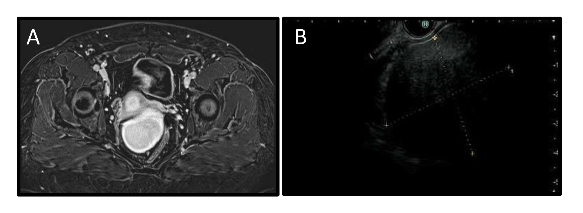
Figure: Figure A: Pelvic MRI demonstrating an enhancing mass arising from the posterior uterine body
Figure B: Rectal EUS showing a 6.6 x5.6 cm hypoechoic mass in the cul-de-sac between the sigmoid colon and the uterus
Figure B: Rectal EUS showing a 6.6 x5.6 cm hypoechoic mass in the cul-de-sac between the sigmoid colon and the uterus
Disclosures:
Jayashrei Sairam indicated no relevant financial relationships.
Akshay Duddu indicated no relevant financial relationships.
Rahul Sharma indicated no relevant financial relationships.
Neeraj Parkash indicated no relevant financial relationships.
Niraj Jani indicated no relevant financial relationships.
Jayashrei Sairam, MD, Akshay Duddu, MD, Rahul Sharma, MD, Neeraj Parkash, MD, Niraj Jani, MD. P2421 - Solitary Fibrous Tumor of the Pelvis Mimicking a Uterine Leiomyoma, ACG 2024 Annual Scientific Meeting Abstracts. Philadelphia, PA: American College of Gastroenterology.

