Monday Poster Session
Category: GI Bleeding
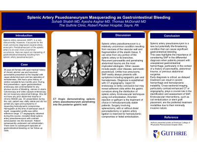
P2486 - Splenic Artery Pseudoaneurysm Masquerading as Gastrointestinal Bleeding
Monday, October 28, 2024
10:30 AM - 4:00 PM ET
Location: Exhibit Hall E

Has Audio
- SS
Sohaib Shabih, MD
Guthrie Robert Packer Hospital
Sayre, PA
Presenting Author(s)
Sohaib Shabih, MD
Guthrie Robert Packer Hospital, Sayre, PA
Introduction: Splenic artery aneurysm is a rare clinical entity. However, it accounts for the most commonly diagnosed visceral artery aneurysm. Pseudoaneurysm of the splenic artery is an even more uncommon occurrence. Here we report an interesting of gastrointestinal bleeding resulting from splenic artery pseudoaneurysm.
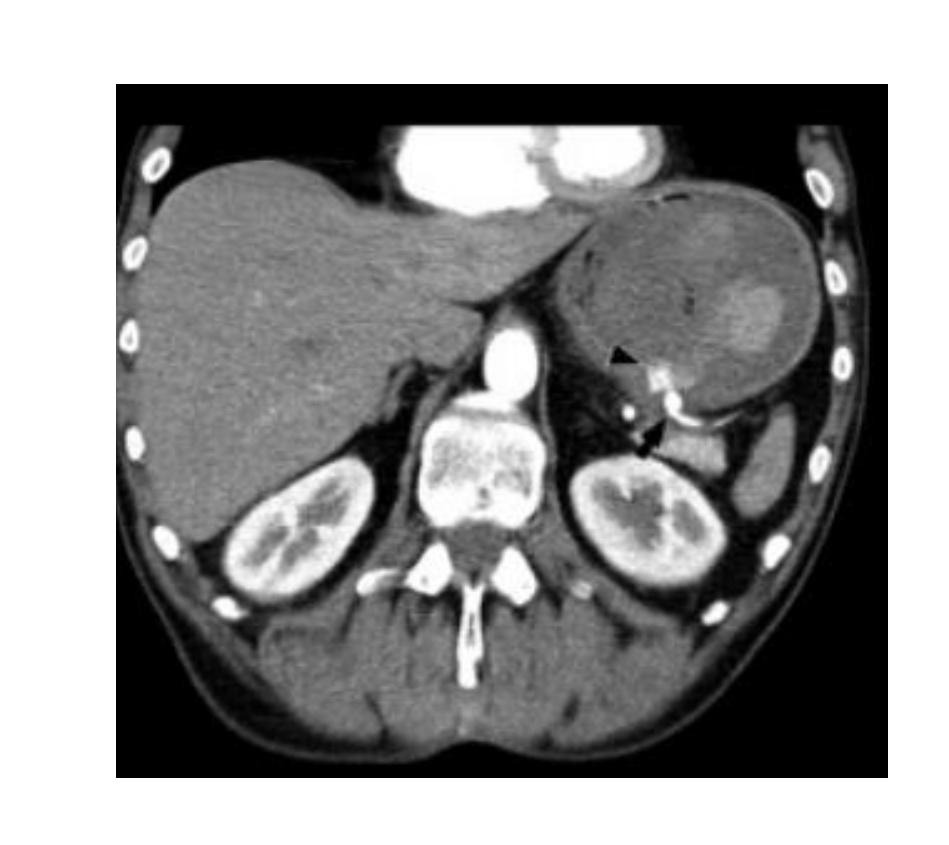
Case Description/Methods: 36 years old female with past medical history of chronic alcohol use, recurrent pancreatitis presented to the hospital with vague abdominal pain and two episodes of hematemesis. She had a prior history of similar complaints two months ago. During that presentation, upper gastrointestinal endoscopy was unremarkable for any obvious source of bleeding,varices or ulcers. Colonoscopy and video capsule enteroscopy did not reveal any abnormal findings.She did not have any prior abdominal trauma or chronic NSAID use.On presentation during this visit, patient was vitally stable and did not exhibit any signs and symptoms of impending hemodynamic compromise. Hemoglobin at the time of admission was 6 mg/dl, coagulation profile was within refrence range. CT angiography obtained during this course, revealed distal splenic artery pseudoaneurysm with contrast extravasation into the stomach. Patient subsequently underwent coil embolization. There were no subsequent episodes of gastrointestinal bleeding on her follow up visits.
Discussion: Splenic artery pseudoaneurysm (SAP) is a relatively uncommon condition resulting from necrosis of the vascular wall and fragmantation of the elastic tissue. It can arise from any portion of the splenic artery or its branches. Recurrent pancreatitis and penetrating abdominal trauma are the most implicated etiologies. Other causes include peptic ulcer disease, pancreatic psuedocyst. Unlike true aneurysms, SAP nearly always presents with symptoms including epigastric pain and hematemesis. Diagnosis is established with CT angiography. Upper GI endoscopy is rarely conclusive but may reveal adherent clots within the gastric curvature along the distribution of splenic artery. Endovascular techniques using coil embolization,thrombin injection or gelfoam is the treatment of choice in hemodynamically stable patients. Surgery involving splenectomy, with or without distal pancreatectomy or splenic artery ligation is reserved for hemodynamic compromise or failed embolization.

Disclosures:
Sohaib Shabih, MD. P2486 - Splenic Artery Pseudoaneurysm Masquerading as Gastrointestinal Bleeding, ACG 2024 Annual Scientific Meeting Abstracts. Philadelphia, PA: American College of Gastroenterology.
Guthrie Robert Packer Hospital, Sayre, PA
Introduction: Splenic artery aneurysm is a rare clinical entity. However, it accounts for the most commonly diagnosed visceral artery aneurysm. Pseudoaneurysm of the splenic artery is an even more uncommon occurrence. Here we report an interesting of gastrointestinal bleeding resulting from splenic artery pseudoaneurysm.
Case Description/Methods: 36 years old female with past medical history of chronic alcohol use, recurrent pancreatitis presented to the hospital with vague abdominal pain and two episodes of hematemesis. She had a prior history of similar complaints two months ago. During that presentation, upper gastrointestinal endoscopy was unremarkable for any obvious source of bleeding,varices or ulcers. Colonoscopy and video capsule enteroscopy did not reveal any abnormal findings.She did not have any prior abdominal trauma or chronic NSAID use.On presentation during this visit, patient was vitally stable and did not exhibit any signs and symptoms of impending hemodynamic compromise. Hemoglobin at the time of admission was 6 mg/dl, coagulation profile was within refrence range. CT angiography obtained during this course, revealed distal splenic artery pseudoaneurysm with contrast extravasation into the stomach. Patient subsequently underwent coil embolization. There were no subsequent episodes of gastrointestinal bleeding on her follow up visits.
Discussion: Splenic artery pseudoaneurysm (SAP) is a relatively uncommon condition resulting from necrosis of the vascular wall and fragmantation of the elastic tissue. It can arise from any portion of the splenic artery or its branches. Recurrent pancreatitis and penetrating abdominal trauma are the most implicated etiologies. Other causes include peptic ulcer disease, pancreatic psuedocyst. Unlike true aneurysms, SAP nearly always presents with symptoms including epigastric pain and hematemesis. Diagnosis is established with CT angiography. Upper GI endoscopy is rarely conclusive but may reveal adherent clots within the gastric curvature along the distribution of splenic artery. Endovascular techniques using coil embolization,thrombin injection or gelfoam is the treatment of choice in hemodynamically stable patients. Surgery involving splenectomy, with or without distal pancreatectomy or splenic artery ligation is reserved for hemodynamic compromise or failed embolization.

Figure: CT angio revealing splenic artery pseudoaneurysm penetrating the posterior gastric wall
Disclosures:
Sohaib Shabih indicated no relevant financial relationships.
Sohaib Shabih, MD. P2486 - Splenic Artery Pseudoaneurysm Masquerading as Gastrointestinal Bleeding, ACG 2024 Annual Scientific Meeting Abstracts. Philadelphia, PA: American College of Gastroenterology.
