Monday Poster Session
Category: GI Bleeding
P2503 - An Unusual Case of Bleeding Infectious Duodenal Mass
Monday, October 28, 2024
10:30 AM - 4:00 PM ET
Location: Exhibit Hall E

Has Audio
- AV
Aditya Vyas, MD
Louisiana State University Health
Shreveport, LA
Presenting Author(s)
Aditya Vyas, MD1, Dhruvkumar Patel, MBBS2, Karan Sachdeva, MD3, Rohit Goyal, MD4, Ross Dies, MD3, Kshitij Arora, MD1, Hassaan A. Zia, MD3
1Louisiana State University Health, Shreveport, LA; 2LSU Health Science Center, Shreveport, LA; 3LSU Health, Shreveport, LA; 4Ochsner LSU Health, Shreveport, LA
Introduction: Intestinal Histoplasmosis is a rare infection caused by Histoplasma Capsulatum that affects the GI tract usually the terminal ileum or colon.We present a rare case of duodenal mass which was eventually found to be histoplasma infection.
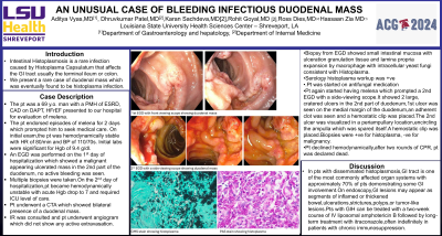
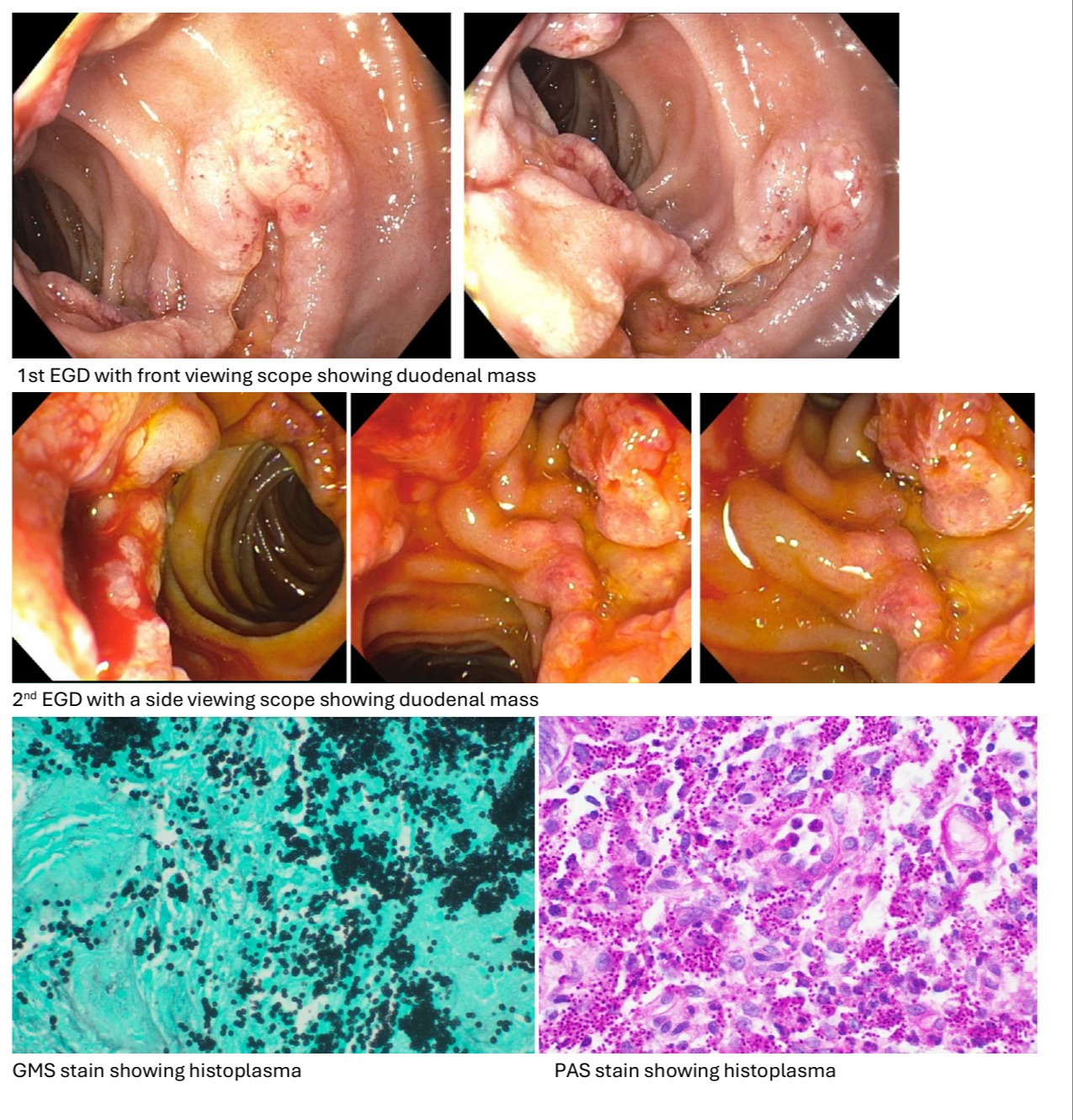
Case Description/Methods: The pt was a 69 y.o. man with a PMH of ESRD, CAD on DAPT, HFrEF presented to our hospital for evaluation of melena. The pt endorsed episodes of melena for 2 days which prompted him to seek medical care. On initial exam,the pt was hemodynamically stable with HR of 80/min and BP of 110/70s. Initial labs were significant for Hgb of 9.4 g/dl.An EGD was performed on the 1st day of hospitalization which showed a malignant appearing ulcerated mass in the 2nd part of the duodenum, no active bleeding was seen.Multiple biopsies were taken.On the 2nd day of hospitalization,pt became hemodynamically unstable with acute Hgb drop to 7 and required ICU level of care.Pt underwent a CTA which showed bilateral presence of a duodenal mass.Due to hemodynamic instability secondary to GI bleed,IR was consulted and pt underwent an angiogram which did not show any active extravasation.Biopsy from EGD showed small intestinal mucosa with ulceration granulation tissue and lamina propria expansion by macrophage with intracellular yeast fungi consistent with Histoplasma. Serology histoplasma workup was +ve for both antigen and antibody.Pt was started on antifungal medication after ID consultation.Pt again started having episodes of melena which prompted a 2nd EGD with a side-viewing scope.It showed 2 large, cratered ulcers in the 2nd part of the duodenum,1st ulcer was seen on the medial margin of the duodenum,an adherent clot was seen and a hemostatic clip was placed.The 2nd ulcer was visualized in a periampullary location,encircling the ampulla which was spared itself.A hemostatic clip was placed.Biopsies were +ve for histoplasma, -ve for malignancy.
Pt declined hemodynamically and CPR was initiated as he was in cardiac arrest. After two rounds of CPR, pt was declared dead.
Discussion: In pts with disseminated histoplasmosis,GI tract is one of the most commonly affected organ systems with approximately 70% of pts demonstrating some GI involvement.On endoscopy,GI lesions may appear as segments of inflamed or thickened bowel,ulcerations,strictures,polyps,or tumor-like lesions.Pts with GIH can be treated with a two-week course of IV liposomal amphotericin B followed by long-term treatment with itraconazole,often indefinitely in patients with chronic immunosuppression.

Disclosures:
Aditya Vyas, MD1, Dhruvkumar Patel, MBBS2, Karan Sachdeva, MD3, Rohit Goyal, MD4, Ross Dies, MD3, Kshitij Arora, MD1, Hassaan A. Zia, MD3. P2503 - An Unusual Case of Bleeding Infectious Duodenal Mass, ACG 2024 Annual Scientific Meeting Abstracts. Philadelphia, PA: American College of Gastroenterology.
1Louisiana State University Health, Shreveport, LA; 2LSU Health Science Center, Shreveport, LA; 3LSU Health, Shreveport, LA; 4Ochsner LSU Health, Shreveport, LA
Introduction: Intestinal Histoplasmosis is a rare infection caused by Histoplasma Capsulatum that affects the GI tract usually the terminal ileum or colon.We present a rare case of duodenal mass which was eventually found to be histoplasma infection.
Case Description/Methods: The pt was a 69 y.o. man with a PMH of ESRD, CAD on DAPT, HFrEF presented to our hospital for evaluation of melena. The pt endorsed episodes of melena for 2 days which prompted him to seek medical care. On initial exam,the pt was hemodynamically stable with HR of 80/min and BP of 110/70s. Initial labs were significant for Hgb of 9.4 g/dl.An EGD was performed on the 1st day of hospitalization which showed a malignant appearing ulcerated mass in the 2nd part of the duodenum, no active bleeding was seen.Multiple biopsies were taken.On the 2nd day of hospitalization,pt became hemodynamically unstable with acute Hgb drop to 7 and required ICU level of care.Pt underwent a CTA which showed bilateral presence of a duodenal mass.Due to hemodynamic instability secondary to GI bleed,IR was consulted and pt underwent an angiogram which did not show any active extravasation.Biopsy from EGD showed small intestinal mucosa with ulceration granulation tissue and lamina propria expansion by macrophage with intracellular yeast fungi consistent with Histoplasma. Serology histoplasma workup was +ve for both antigen and antibody.Pt was started on antifungal medication after ID consultation.Pt again started having episodes of melena which prompted a 2nd EGD with a side-viewing scope.It showed 2 large, cratered ulcers in the 2nd part of the duodenum,1st ulcer was seen on the medial margin of the duodenum,an adherent clot was seen and a hemostatic clip was placed.The 2nd ulcer was visualized in a periampullary location,encircling the ampulla which was spared itself.A hemostatic clip was placed.Biopsies were +ve for histoplasma, -ve for malignancy.
Pt declined hemodynamically and CPR was initiated as he was in cardiac arrest. After two rounds of CPR, pt was declared dead.
Discussion: In pts with disseminated histoplasmosis,GI tract is one of the most commonly affected organ systems with approximately 70% of pts demonstrating some GI involvement.On endoscopy,GI lesions may appear as segments of inflamed or thickened bowel,ulcerations,strictures,polyps,or tumor-like lesions.Pts with GIH can be treated with a two-week course of IV liposomal amphotericin B followed by long-term treatment with itraconazole,often indefinitely in patients with chronic immunosuppression.

Figure: Images from endoscopy and pathology slides
Disclosures:
Aditya Vyas indicated no relevant financial relationships.
Dhruvkumar Patel indicated no relevant financial relationships.
Karan Sachdeva indicated no relevant financial relationships.
Rohit Goyal indicated no relevant financial relationships.
Ross Dies indicated no relevant financial relationships.
Kshitij Arora indicated no relevant financial relationships.
Hassaan A. Zia indicated no relevant financial relationships.
Aditya Vyas, MD1, Dhruvkumar Patel, MBBS2, Karan Sachdeva, MD3, Rohit Goyal, MD4, Ross Dies, MD3, Kshitij Arora, MD1, Hassaan A. Zia, MD3. P2503 - An Unusual Case of Bleeding Infectious Duodenal Mass, ACG 2024 Annual Scientific Meeting Abstracts. Philadelphia, PA: American College of Gastroenterology.
