Monday Poster Session
Category: Interventional Endoscopy
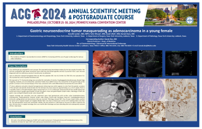
P2810 - Gastric Neuroendocrine Tumor Masquerading as Adenocarcinoma in a Young Female
Monday, October 28, 2024
10:30 AM - 4:00 PM ET
Location: Exhibit Hall E

Has Audio
- GL
Gandhi Lanke, MD
Texas Tech University Health Sciences Center
Lubbock, TX
Presenting Author(s)
Gandhi Lanke, MD, Kanak Das, MD, Tyler Mouw, MD, Asad Ullah, MD
Texas Tech University Health Sciences Center, Lubbock, TX
Introduction: The incidence of Gastric neuroendocrine tumors (GNET) is increasing with the use of upper endoscopy for various other indications.
Case Description/Methods: 23 yr old female was admitted for near syncope. She reported having intermittent melena for the last couple of months. She also has constipation with bowel movement every other day. Decreased appetite and lost 15 pounds in last 1 year. She was diagnosed with iron deficiency anemia 6 months prior to admission.
Labs on admission showed hemoglobin (Hb) 5.8, MCV 63, platelets 565. Iron 10, ferritin 3.0, TIBC 533, Iron saturation 5.0. she received two units of packed red blood cells.
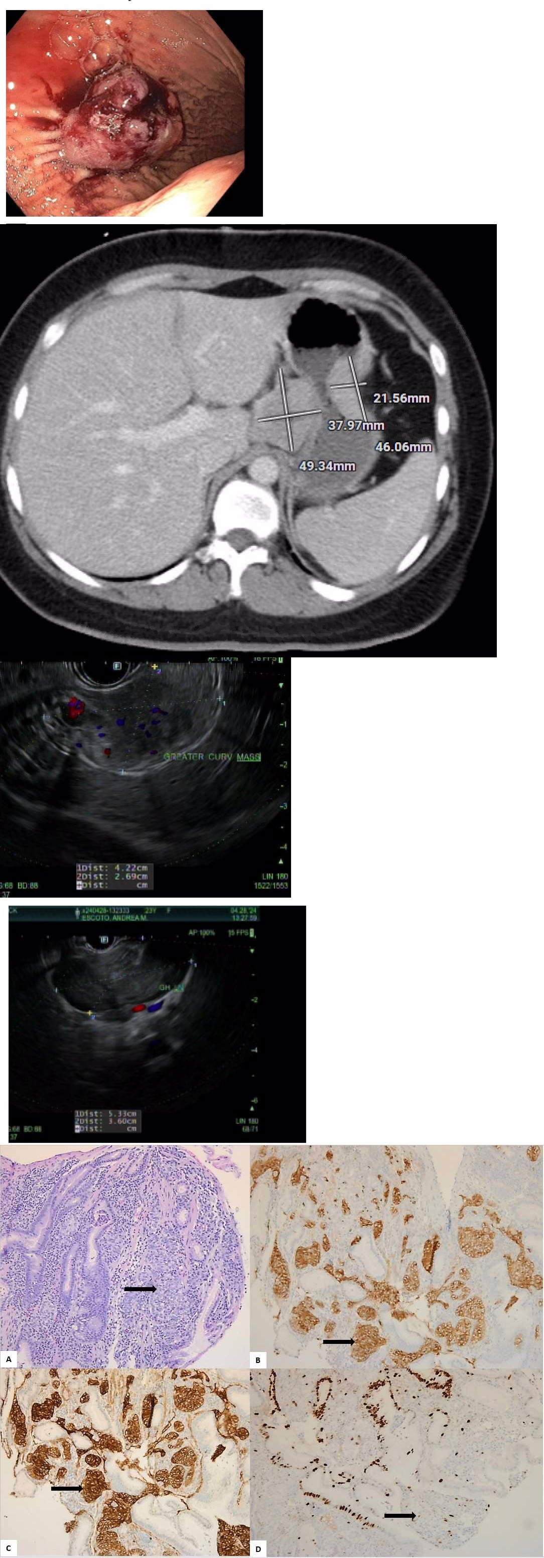
Hb improved to 7.9. Gastroenterology was consulted for evaluation of anemia. Esophagogastroduodenoscopy showed large fungating, localized, non-circumferential, ulcerated, friable mass (20X10 mm) in the mid-portion of the gastric body at the greater curvature of the stomach.Biopsy of the mass consistent with a well differentiated neuroendocrine tumor (NET).
CT chest, abdomen and pelvis showed homogeneously enhancing mass which appears to arise from the greater curvature of the stomach and extends intraluminally into the stomach; the mass measures approximately 4.1 x 1.8 cm axially. There is a masslike cluster in the gastrohepatic region measuring 4.0 x 3.3 cm.Endoscopic ultrasound (EUS) Fine needle biopsy was performed using 22 guage aquireneedle of both the gastric mass and gastrohepatic lymph nodes. Pathology of both the gastric mass and gastrohepatic lymph node showed NET.
Surgical oncology was consulted, and she underwent open total gastrectomy with en-bloc celiac lymphadenectomy, omentectomy, cholecystectomy, ileocolic lymph node biopsy. End-to-side handsewn Roux-en-Y esophagojejunostomy (retrocolic configuration) and Jejunostomy tube was inserted. Pathology showed NET. One of seven lymph nodes, positive for tumor (1/7). Surgical margins are negative Mitotic Rate: Less than 2 mitoses per 2 mm2. Ki-67 Labeling Index: Less than 3%. The postoperative course was uneventful. Initially, she was started on J tube feeds as she was not able to tolerate oral diet. She was seen in surgical oncology clinic one month after discharge and was tolerating diet and subsequently weaned off the J tube feeds.
Discussion: EUS aids in the definitive diagnosis of GNET with nodal involvement. Total gastrectomy with lymphadenectomy is the treatment of choice for Type 3 NET with submucosal involvement and size >2 cm.

Disclosures:
Gandhi Lanke, MD, Kanak Das, MD, Tyler Mouw, MD, Asad Ullah, MD. P2810 - Gastric Neuroendocrine Tumor Masquerading as Adenocarcinoma in a Young Female, ACG 2024 Annual Scientific Meeting Abstracts. Philadelphia, PA: American College of Gastroenterology.
Texas Tech University Health Sciences Center, Lubbock, TX
Introduction: The incidence of Gastric neuroendocrine tumors (GNET) is increasing with the use of upper endoscopy for various other indications.
Case Description/Methods: 23 yr old female was admitted for near syncope. She reported having intermittent melena for the last couple of months. She also has constipation with bowel movement every other day. Decreased appetite and lost 15 pounds in last 1 year. She was diagnosed with iron deficiency anemia 6 months prior to admission.
Labs on admission showed hemoglobin (Hb) 5.8, MCV 63, platelets 565. Iron 10, ferritin 3.0, TIBC 533, Iron saturation 5.0. she received two units of packed red blood cells.
Hb improved to 7.9. Gastroenterology was consulted for evaluation of anemia. Esophagogastroduodenoscopy showed large fungating, localized, non-circumferential, ulcerated, friable mass (20X10 mm) in the mid-portion of the gastric body at the greater curvature of the stomach.Biopsy of the mass consistent with a well differentiated neuroendocrine tumor (NET).
CT chest, abdomen and pelvis showed homogeneously enhancing mass which appears to arise from the greater curvature of the stomach and extends intraluminally into the stomach; the mass measures approximately 4.1 x 1.8 cm axially. There is a masslike cluster in the gastrohepatic region measuring 4.0 x 3.3 cm.Endoscopic ultrasound (EUS) Fine needle biopsy was performed using 22 guage aquireneedle of both the gastric mass and gastrohepatic lymph nodes. Pathology of both the gastric mass and gastrohepatic lymph node showed NET.
Surgical oncology was consulted, and she underwent open total gastrectomy with en-bloc celiac lymphadenectomy, omentectomy, cholecystectomy, ileocolic lymph node biopsy. End-to-side handsewn Roux-en-Y esophagojejunostomy (retrocolic configuration) and Jejunostomy tube was inserted. Pathology showed NET. One of seven lymph nodes, positive for tumor (1/7). Surgical margins are negative Mitotic Rate: Less than 2 mitoses per 2 mm2. Ki-67 Labeling Index: Less than 3%. The postoperative course was uneventful. Initially, she was started on J tube feeds as she was not able to tolerate oral diet. She was seen in surgical oncology clinic one month after discharge and was tolerating diet and subsequently weaned off the J tube feeds.
Discussion: EUS aids in the definitive diagnosis of GNET with nodal involvement. Total gastrectomy with lymphadenectomy is the treatment of choice for Type 3 NET with submucosal involvement and size >2 cm.

Figure: NET IMAGES
Disclosures:
Gandhi Lanke indicated no relevant financial relationships.
Kanak Das indicated no relevant financial relationships.
Tyler Mouw indicated no relevant financial relationships.
Asad Ullah indicated no relevant financial relationships.
Gandhi Lanke, MD, Kanak Das, MD, Tyler Mouw, MD, Asad Ullah, MD. P2810 - Gastric Neuroendocrine Tumor Masquerading as Adenocarcinoma in a Young Female, ACG 2024 Annual Scientific Meeting Abstracts. Philadelphia, PA: American College of Gastroenterology.
