Monday Poster Session
Category: Interventional Endoscopy
P2823 - Endoscopic Diagnosis of Extranodal Diffuse Large B-Cell Lymphoma With Gastric and Pancreatic Involvement
Monday, October 28, 2024
10:30 AM - 4:00 PM ET
Location: Exhibit Hall E

Has Audio
- RB
Rishi Bolla, MD
University of South Florida Morsani College of Medicine
Tampa, FL
Presenting Author(s)
Rishi Bolla, MD1, Sonya Bhaskar, MD2, Gino Piparo, MD3, Prasad Kulkarni, MD2
1University of South Florida Morsani College of Medicine, Tampa, FL; 2University of South Florida, Tampa, FL; 3James A. Haley Veterans' Hospital, Tampa, FL
Introduction: Gastrointestinal manifestation of diffuse large B-cell lymphoma (DLBCL) is a relatively rare phenomenon. Patients commonly experience nonspecific upper GI symptoms including pain, indigestion, nausea/vomiting, or early satiety (1). The most common site involves the stomach, and endoscopically it can present as ulceration, wall thickening, or gastric polyps (1,2). Here we present a case where esophagogastroduodenoscopy (EGD) and endoscopic ultrasound (EUS) were successfully used to diagnose DLBCL with significant extranodal involvement.
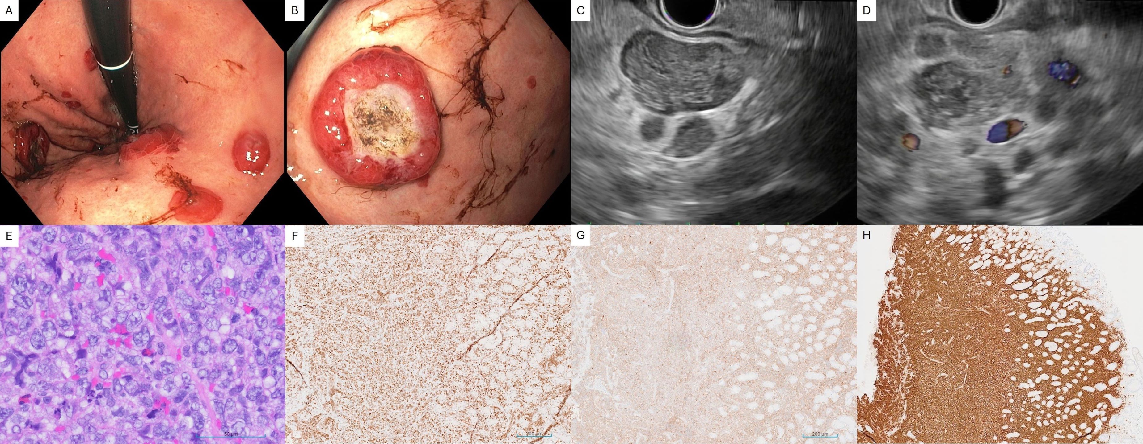
Case Description/Methods: A 74-year-old male with four-month history of intractable hiccups and progressive, debilitating right hip pain presented to our institution after outpatient magnetic resonance imaging revealed metastatic lesion in the right iliac bone. Further imaging identified additional osseous lesions, lymphadenopathy, and a mass in the mid-body of the pancreas. EGD unexpectedly revealed multiple nodules between 6mm and 2.5cm in the stomach and duodenum. EUS revealed enlarged celiac lymph nodes and a 3.4cm x 2.6cm lesion in the pancreatic tail. Fine needle aspiration and cytology of the pancreatic mass was inconclusive, however biopsies of the gastric nodule and celiac lymph node revealed CD10+ lymphoproliferative B-cells consistent with DLBCL. Additional biopsy of the right hip lesion and axillary lymph node further confirmed the diagnosis.
Discussion: Since our patient had Stage IV disease at the time of presentation, it is difficult to determine per criteria if he had primary gastric DLBCL or if he had metastasis to the stomach (3). The initial plan was to use EUS to obtain biopsies of the pancreatic mass, but the EGD biopsies of the gastric nodules yielded the diagnosis. While EUS can identify suspicious peri-gastrointestinal lymph nodes and submucosal disease in GI lymphoma, EGD has also been shown to have high diagnostic accuracy (1). Our case demonstrates these qualities and highlights the utility of endoscopy in establishing the diagnosis in this fairly uncommon disease presentation.
References
1. Shirwaikar et al. Gastrointestinal lymphoma: the new mimic. BMJ Open Gastroenterol. 2019;6(1):e000320. Published 2019 Sep 13.
2. Bai Z, Zhou Y. A systematic review of primary gastric diffuse large B-cell lymphoma: Clinical diagnosis, staging, treatment and prognostic factors. Leuk Res. 2021;111:106716.
3. Suresh et al. Primary gastrointestinal diffuse large B-cell lymphoma: A prospective study from South India. South Asian J Cancer. 2019;8(1):57-59.

Disclosures:
Rishi Bolla, MD1, Sonya Bhaskar, MD2, Gino Piparo, MD3, Prasad Kulkarni, MD2. P2823 - Endoscopic Diagnosis of Extranodal Diffuse Large B-Cell Lymphoma With Gastric and Pancreatic Involvement, ACG 2024 Annual Scientific Meeting Abstracts. Philadelphia, PA: American College of Gastroenterology.
1University of South Florida Morsani College of Medicine, Tampa, FL; 2University of South Florida, Tampa, FL; 3James A. Haley Veterans' Hospital, Tampa, FL
Introduction: Gastrointestinal manifestation of diffuse large B-cell lymphoma (DLBCL) is a relatively rare phenomenon. Patients commonly experience nonspecific upper GI symptoms including pain, indigestion, nausea/vomiting, or early satiety (1). The most common site involves the stomach, and endoscopically it can present as ulceration, wall thickening, or gastric polyps (1,2). Here we present a case where esophagogastroduodenoscopy (EGD) and endoscopic ultrasound (EUS) were successfully used to diagnose DLBCL with significant extranodal involvement.
Case Description/Methods: A 74-year-old male with four-month history of intractable hiccups and progressive, debilitating right hip pain presented to our institution after outpatient magnetic resonance imaging revealed metastatic lesion in the right iliac bone. Further imaging identified additional osseous lesions, lymphadenopathy, and a mass in the mid-body of the pancreas. EGD unexpectedly revealed multiple nodules between 6mm and 2.5cm in the stomach and duodenum. EUS revealed enlarged celiac lymph nodes and a 3.4cm x 2.6cm lesion in the pancreatic tail. Fine needle aspiration and cytology of the pancreatic mass was inconclusive, however biopsies of the gastric nodule and celiac lymph node revealed CD10+ lymphoproliferative B-cells consistent with DLBCL. Additional biopsy of the right hip lesion and axillary lymph node further confirmed the diagnosis.
Discussion: Since our patient had Stage IV disease at the time of presentation, it is difficult to determine per criteria if he had primary gastric DLBCL or if he had metastasis to the stomach (3). The initial plan was to use EUS to obtain biopsies of the pancreatic mass, but the EGD biopsies of the gastric nodules yielded the diagnosis. While EUS can identify suspicious peri-gastrointestinal lymph nodes and submucosal disease in GI lymphoma, EGD has also been shown to have high diagnostic accuracy (1). Our case demonstrates these qualities and highlights the utility of endoscopy in establishing the diagnosis in this fairly uncommon disease presentation.
References
1. Shirwaikar et al. Gastrointestinal lymphoma: the new mimic. BMJ Open Gastroenterol. 2019;6(1):e000320. Published 2019 Sep 13.
2. Bai Z, Zhou Y. A systematic review of primary gastric diffuse large B-cell lymphoma: Clinical diagnosis, staging, treatment and prognostic factors. Leuk Res. 2021;111:106716.
3. Suresh et al. Primary gastrointestinal diffuse large B-cell lymphoma: A prospective study from South India. South Asian J Cancer. 2019;8(1):57-59.

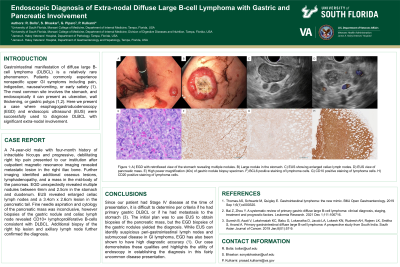
Figure: Figure 1: A) EGD with retroflexed view of the stomach revealing multiple nodules. B) Large nodule in the stomach. C) EUS showing enlarged celiac lymph nodes. D) EUS view of pancreatic mass. E) High power magnification (40x) of gastric nodule biopsy specimen. F) BCL6 positive staining of lymphoma cells. G) CD10 positive staining of lymphoma cells. H) CD20 positive staining of lymphoma cells.
Disclosures:
Rishi Bolla indicated no relevant financial relationships.
Sonya Bhaskar indicated no relevant financial relationships.
Gino Piparo indicated no relevant financial relationships.
Prasad Kulkarni indicated no relevant financial relationships.
Rishi Bolla, MD1, Sonya Bhaskar, MD2, Gino Piparo, MD3, Prasad Kulkarni, MD2. P2823 - Endoscopic Diagnosis of Extranodal Diffuse Large B-Cell Lymphoma With Gastric and Pancreatic Involvement, ACG 2024 Annual Scientific Meeting Abstracts. Philadelphia, PA: American College of Gastroenterology.
