Monday Poster Session
Category: Interventional Endoscopy
P2829 - Endoscopic Ultrasound-Directed Transgastric FNA in Diagnosis of Late Metastasis of Renal Cell Carcinoma
Monday, October 28, 2024
10:30 AM - 4:00 PM ET
Location: Exhibit Hall E

Has Audio
- LD
Luke Durbin, MD
Carilion Clinic
Roanoke, VA
Presenting Author(s)
Luke Durbin, MD, William Abel, MD, Joel Joseph, MD, Adil Mir, MD, Seemeen Hassan, MD, Varun Kesar, MD, Vivek Kesar, MD
Carilion Clinic, Roanoke, VA
Introduction: Renal cell carcinoma (RCC) is an aggressive malignancy that most commonly metastasizes to the lung, bone, and lymph nodes with late metastases also possible years after radical nephrectomy. Biopsy of a lesion suspicious for metastasis is ideal for diagnosis but the increasingly common Roux-en-Y phenotype poses a challenge in patients requiring fine needle aspiration (FNA) of the head of pancreas. Endoscopic ultrasound-directed transgastric FNA procedure provides a minimally invasive option in this subset. We detail a care of late RCC metastasis to the head of pancreas diagnosed with endoscopic ultrasound-directed transgastric FNA.
Case Description/Methods: A 48-year-old female with history of Roux-en-Y gastric bypass and left renal cell carcinoma treated with nephrectomy seven years prior presented to the primary care clinic with complaint of right-sided abdominal pain. MRI demonstrated a 3.4 x 3.2 x 3.7 cm rounded intermediate hyperintense T2 signal lesion in the downstream pancreatic neck and head with attenuation of the pancreatic duct, initially concerning for neuroendocrine neoplasm. Dotatate scan was denied by insurance. The patient underwent EUS guided gastro-gastric fistula creation via a 20 mm x 10 mm cautery enhanced AxiosTM. Transgastric endoscopic ultrasound-guided FNA of the head of pancreas was attained. Pathology resulted in malignant cells compatible with metastatic renal cell carcinoma. By immunocytochemistry, the biopsied cells exhibited immunoreactivity with AE1/AE3 pancytokeratin, PAX-8 and RCC. CD10 was positive but CK7, Villin and Synaptophysin were negative, the same chemical profile as the previous renal cell carcinoma, confirming late metastasis. The patient was subsequently referred to medical oncology for palliative treatment.
Discussion: Renal cell carcinoma is notorious for its highly aggressive nature and tendency to metastasize, even following radical nephrectomy. To our knowledge, there are no cases in the literature that report the diagnosis of pancreatic renal cell carcinoma metastasis by way of endoscopic ultrasound-directed transgastric FNA. In this patient’s case, the other diagnostic alternative that was being considered was a Whipple procedure, as the mass was initially presumed to be a neuroendocrine tumor. Endoscopic ultrasound-directed transgastric FNA in this instance allowed diagnosis of metastatic renal cell carcinoma and spared the patient a radical surgery that was not indicated.

Disclosures:
Luke Durbin, MD, William Abel, MD, Joel Joseph, MD, Adil Mir, MD, Seemeen Hassan, MD, Varun Kesar, MD, Vivek Kesar, MD. P2829 - Endoscopic Ultrasound-Directed Transgastric FNA in Diagnosis of Late Metastasis of Renal Cell Carcinoma, ACG 2024 Annual Scientific Meeting Abstracts. Philadelphia, PA: American College of Gastroenterology.
Carilion Clinic, Roanoke, VA
Introduction: Renal cell carcinoma (RCC) is an aggressive malignancy that most commonly metastasizes to the lung, bone, and lymph nodes with late metastases also possible years after radical nephrectomy. Biopsy of a lesion suspicious for metastasis is ideal for diagnosis but the increasingly common Roux-en-Y phenotype poses a challenge in patients requiring fine needle aspiration (FNA) of the head of pancreas. Endoscopic ultrasound-directed transgastric FNA procedure provides a minimally invasive option in this subset. We detail a care of late RCC metastasis to the head of pancreas diagnosed with endoscopic ultrasound-directed transgastric FNA.
Case Description/Methods: A 48-year-old female with history of Roux-en-Y gastric bypass and left renal cell carcinoma treated with nephrectomy seven years prior presented to the primary care clinic with complaint of right-sided abdominal pain. MRI demonstrated a 3.4 x 3.2 x 3.7 cm rounded intermediate hyperintense T2 signal lesion in the downstream pancreatic neck and head with attenuation of the pancreatic duct, initially concerning for neuroendocrine neoplasm. Dotatate scan was denied by insurance. The patient underwent EUS guided gastro-gastric fistula creation via a 20 mm x 10 mm cautery enhanced AxiosTM. Transgastric endoscopic ultrasound-guided FNA of the head of pancreas was attained. Pathology resulted in malignant cells compatible with metastatic renal cell carcinoma. By immunocytochemistry, the biopsied cells exhibited immunoreactivity with AE1/AE3 pancytokeratin, PAX-8 and RCC. CD10 was positive but CK7, Villin and Synaptophysin were negative, the same chemical profile as the previous renal cell carcinoma, confirming late metastasis. The patient was subsequently referred to medical oncology for palliative treatment.
Discussion: Renal cell carcinoma is notorious for its highly aggressive nature and tendency to metastasize, even following radical nephrectomy. To our knowledge, there are no cases in the literature that report the diagnosis of pancreatic renal cell carcinoma metastasis by way of endoscopic ultrasound-directed transgastric FNA. In this patient’s case, the other diagnostic alternative that was being considered was a Whipple procedure, as the mass was initially presumed to be a neuroendocrine tumor. Endoscopic ultrasound-directed transgastric FNA in this instance allowed diagnosis of metastatic renal cell carcinoma and spared the patient a radical surgery that was not indicated.

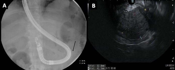
Figure: A. Fluoroscopic image depicting passage of endoscope through lumen apposing stent (denoted by black arrow).
B. Endoscopic ultrasound showing the pancreatic head mass measuring 32.8 mm x 41.5 mm.
B. Endoscopic ultrasound showing the pancreatic head mass measuring 32.8 mm x 41.5 mm.
Disclosures:
Luke Durbin indicated no relevant financial relationships.
William Abel indicated no relevant financial relationships.
Joel Joseph indicated no relevant financial relationships.
Adil Mir indicated no relevant financial relationships.
Seemeen Hassan indicated no relevant financial relationships.
Varun Kesar indicated no relevant financial relationships.
Vivek Kesar indicated no relevant financial relationships.
Luke Durbin, MD, William Abel, MD, Joel Joseph, MD, Adil Mir, MD, Seemeen Hassan, MD, Varun Kesar, MD, Vivek Kesar, MD. P2829 - Endoscopic Ultrasound-Directed Transgastric FNA in Diagnosis of Late Metastasis of Renal Cell Carcinoma, ACG 2024 Annual Scientific Meeting Abstracts. Philadelphia, PA: American College of Gastroenterology.
