Monday Poster Session
Category: Interventional Endoscopy
P2831 - Pancreatic Mass in a Roux-en-Y Patient Requiring Modified EDGE Procedure for EUS Evaluation
Monday, October 28, 2024
10:30 AM - 4:00 PM ET
Location: Exhibit Hall E

Has Audio

Samar Taqvi, DO
Baylor All Saints
Fort Worth, TX
Presenting Author(s)
Daniel Cain, DO, Sidart Pradeep, MD, MS, Samar Taqvi, DO, Melvin Simien, MD
Baylor All Saints, Fort Worth, TX
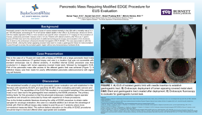
Introduction: Pancreatic cancer is the 3rd most common cause of cancer related mortality in the USA with a mortality rate of 11.1 per 100,000 people, accounting for 7% of all cancer related deaths in the USA [1-3]. endoscopic ultrasound (EUS) with fine needle aspiration (FNA) has been shown to be more sensitive and specific when compared to CT imaging for evaluation of pancreatic masses, especially small pancreatic masses under 2cm [4]. Patients with altered anatomy after Roux-en-Y gastric bypass (RYGB), have led to the development of additional techniques for biopsy and evaluation of GI structures, one of which is the endoscopic ultrasound-directed transgastric ERCP (EDGE). This was expounded on with the Internal EDGE, utilizing the lumen apposing covered metal stent to create a gastrogastric fistula for instrumentation to pass through in order to access the excluded gastric region without any transcutaneous hardware [5,6].
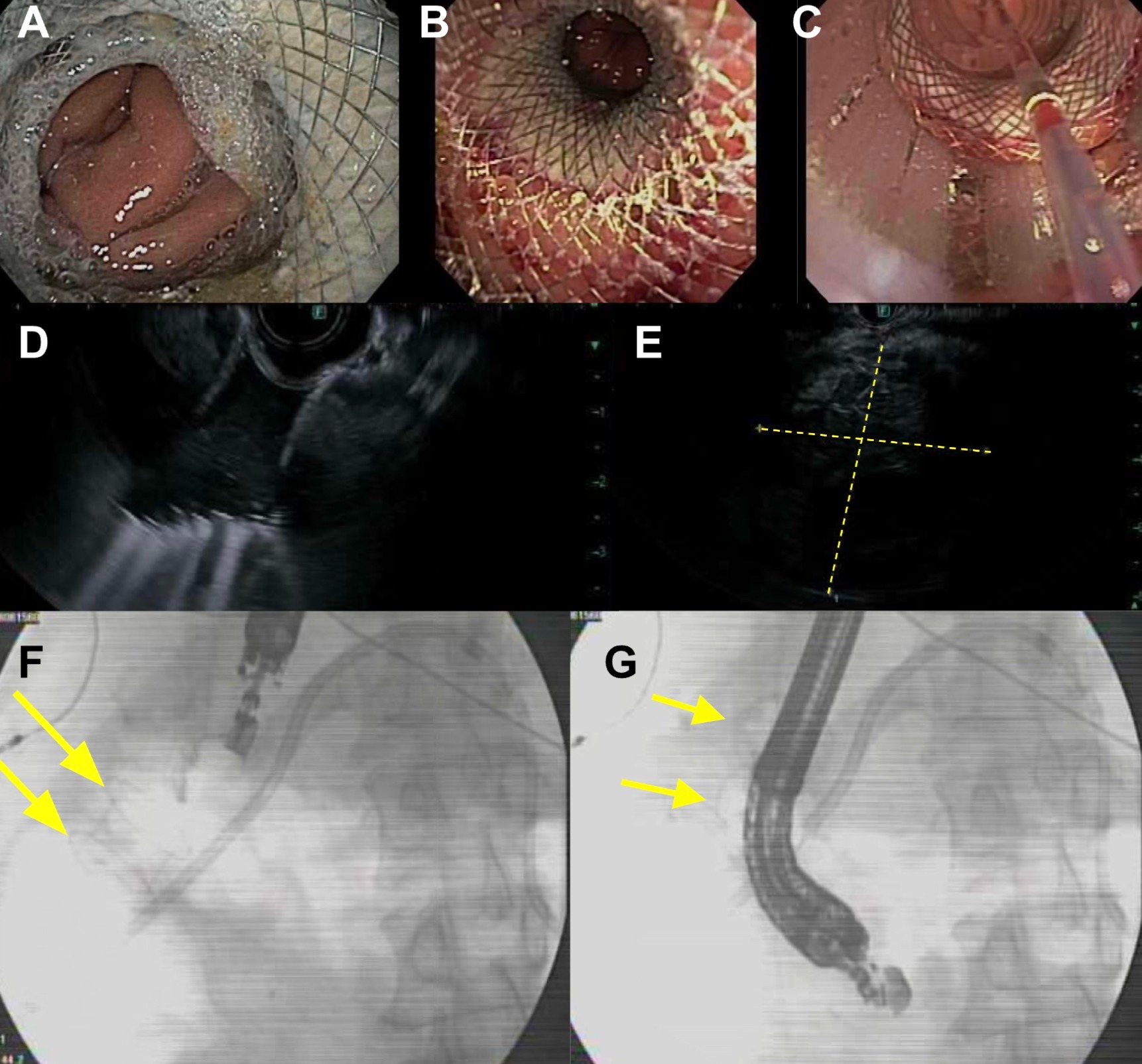
Case Description/Methods: This is a presentation of a modified Internal EDGE procedure in a 70 year old male with RYGB history and large pancreatic mass that was unable to be sampled by transcutaneous CT guided biopsy or EUS alone. A modified Internal EDGE procedure was then conducted in 2 stages with lumen apposing covered metal stent, followed by transgastric EUS-FNA of the pancreatic mass after access to the isolated gastric limb was achieved (Figure 1). A definitive biopsy for pancreatic adenocarcinoma was then able to be made based on this biopsy.
Discussion: The established benefits of using EUS for pancreatic cancer evaluation are well established in the literature with sensitivity 85-92% and specificity 96-98% when evaluating pancreatic cancers and using FNA [9]. The pairing of this imaging modality with biopsy capability is what ultimately allowed for a viable sample to be attained in this case. Using the EDGE procedure to gain access to the isolated gastric pouch allowed for EUS-FNA to be utilized which could not be completed with a double balloon endoscopy (DBE), which has been shown to be another modality for accessing the isolated pouch [7].

Disclosures:
Daniel Cain, DO, Sidart Pradeep, MD, MS, Samar Taqvi, DO, Melvin Simien, MD. P2831 - Pancreatic Mass in a Roux-en-Y Patient Requiring Modified EDGE Procedure for EUS Evaluation, ACG 2024 Annual Scientific Meeting Abstracts. Philadelphia, PA: American College of Gastroenterology.
Baylor All Saints, Fort Worth, TX
Introduction: Pancreatic cancer is the 3rd most common cause of cancer related mortality in the USA with a mortality rate of 11.1 per 100,000 people, accounting for 7% of all cancer related deaths in the USA [1-3]. endoscopic ultrasound (EUS) with fine needle aspiration (FNA) has been shown to be more sensitive and specific when compared to CT imaging for evaluation of pancreatic masses, especially small pancreatic masses under 2cm [4]. Patients with altered anatomy after Roux-en-Y gastric bypass (RYGB), have led to the development of additional techniques for biopsy and evaluation of GI structures, one of which is the endoscopic ultrasound-directed transgastric ERCP (EDGE). This was expounded on with the Internal EDGE, utilizing the lumen apposing covered metal stent to create a gastrogastric fistula for instrumentation to pass through in order to access the excluded gastric region without any transcutaneous hardware [5,6].
Case Description/Methods: This is a presentation of a modified Internal EDGE procedure in a 70 year old male with RYGB history and large pancreatic mass that was unable to be sampled by transcutaneous CT guided biopsy or EUS alone. A modified Internal EDGE procedure was then conducted in 2 stages with lumen apposing covered metal stent, followed by transgastric EUS-FNA of the pancreatic mass after access to the isolated gastric limb was achieved (Figure 1). A definitive biopsy for pancreatic adenocarcinoma was then able to be made based on this biopsy.
Discussion: The established benefits of using EUS for pancreatic cancer evaluation are well established in the literature with sensitivity 85-92% and specificity 96-98% when evaluating pancreatic cancers and using FNA [9]. The pairing of this imaging modality with biopsy capability is what ultimately allowed for a viable sample to be attained in this case. Using the EDGE procedure to gain access to the isolated gastric pouch allowed for EUS-FNA to be utilized which could not be completed with a double balloon endoscopy (DBE), which has been shown to be another modality for accessing the isolated pouch [7].

Figure: FIGURE 1: A&B) Stable gastrogastric tract with stent. C) FNA needle being introduced through gastrogastric tract for pancreatic mass biopsy. D) EUS of gastrogastric stent tract. E) EUS showing pancreatic mass F&G) Fluoroscopy showing endoscope approaching and traversing the gastrogastric tract through lumen apposing metal covered stent.
Disclosures:
Daniel Cain indicated no relevant financial relationships.
Sidart Pradeep indicated no relevant financial relationships.
Samar Taqvi indicated no relevant financial relationships.
Melvin Simien indicated no relevant financial relationships.
Daniel Cain, DO, Sidart Pradeep, MD, MS, Samar Taqvi, DO, Melvin Simien, MD. P2831 - Pancreatic Mass in a Roux-en-Y Patient Requiring Modified EDGE Procedure for EUS Evaluation, ACG 2024 Annual Scientific Meeting Abstracts. Philadelphia, PA: American College of Gastroenterology.
