Monday Poster Session
Category: Interventional Endoscopy
P2834 - A Complex EndoVAC Leak Closure in a Community Hospital
Monday, October 28, 2024
10:30 AM - 4:00 PM ET
Location: Exhibit Hall E

Has Audio

Shalva Eliava, MD
Englewood Hospital and Medical Center
Englewood, NJ
Presenting Author(s)
Shalva Eliava, MD1, Chadane Thompson, MD1, Danielle Pope, MD2, Dipal Patel, MD1, Mitchell Spinnell, MD1
1Englewood Hospital and Medical Center, Englewood, NJ; 2Englewood Hospital and Medical Center, Bogota, NJ
Introduction: Endoscopic vacuum therapy (EVT) is an established method of treating anastomotic leaks in the upper and lower gastrointestinal (GI) tract. It is a safe, effective, and minimally invasive procedure that can avoid the need for more invasive surgical interventions and reduce morbidity and mortality. This technique is usually reserved for academic tertiary centers. We present a case of a gastric leak after bariatric surgery in a community hospital that was successfully treated with EVT.
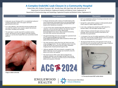
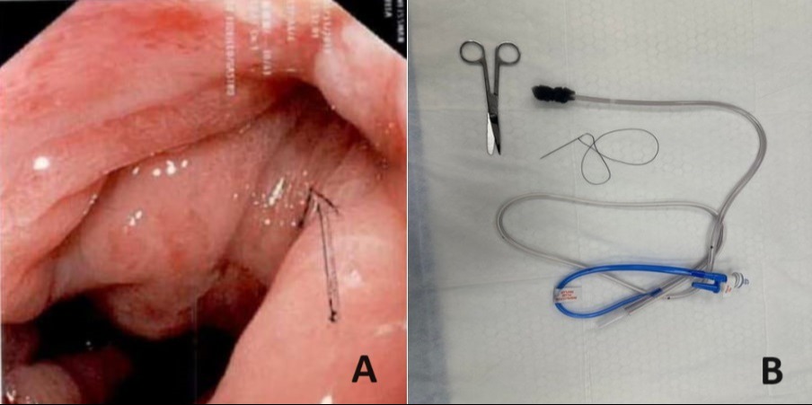
Case Description/Methods: A 78-year-old woman with a history of morbid obesity with recent laparoscopic sleeve gastrectomy and paraesophageal hernia repair presented several weeks postoperatively with dyspnea and malodorous eructation. CT scan of the chest revealed large left subpulmonic air, fluid collection, and oral contrast leak from the proximal sleeve gastrectomy suture line, for which laparoscopic paraesophageal hernia repair, video-assisted thoracotomy, and chest tube insertion were performed. The gastroenterology team was consulted for a persistent leak from the gastroesophageal junction (GEJ). Esophagogastroduodenoscopy (EGD) revealed an 8-9mm open defect at the GEJ (Image A). The initial attempt to divert flow from the fistula was done using esophageal stents. This was ineffective and an over-the-scope clip (OTSC) was used to close the defect. Repeated imaging studies showed persistent gastric leak. EGD was repeated, OTSC was removed, a nasogastric tube (NGT) endoscopic vacuum sponge was inserted to close the defect, and suction was applied. The wound vacuum NGT suction device (Image B) was designed, deployed, and exchanged multiple times during hospitalization. This led to successful closure of the fistulous defect and cessation of the leak.
Discussion: EVT is an established method of treating anastomotic leaks in the upper and lower GI tract. During EVT, a polyurethane sponge is endoscopically placed inside a defect to apply negative pressure. In our case, a sponge was sutured to an NGT, placed in a fistulous defect, and exchanged multiple times. Defect healing is achieved through continuous abscess drainage, which decreases bacterial colonization and promotes tissue granulation. In retrospect, the cost of multiple applications of EVT was less costly than commercially available devices used unsuccessfully in the initial management of this patient. We demonstrated that this novel approach to EVT can be cost-effective and successfully used to close upper GI defects, even in a small, community-based center.

Disclosures:
Shalva Eliava, MD1, Chadane Thompson, MD1, Danielle Pope, MD2, Dipal Patel, MD1, Mitchell Spinnell, MD1. P2834 - A Complex EndoVAC Leak Closure in a Community Hospital, ACG 2024 Annual Scientific Meeting Abstracts. Philadelphia, PA: American College of Gastroenterology.
1Englewood Hospital and Medical Center, Englewood, NJ; 2Englewood Hospital and Medical Center, Bogota, NJ
Introduction: Endoscopic vacuum therapy (EVT) is an established method of treating anastomotic leaks in the upper and lower gastrointestinal (GI) tract. It is a safe, effective, and minimally invasive procedure that can avoid the need for more invasive surgical interventions and reduce morbidity and mortality. This technique is usually reserved for academic tertiary centers. We present a case of a gastric leak after bariatric surgery in a community hospital that was successfully treated with EVT.
Case Description/Methods: A 78-year-old woman with a history of morbid obesity with recent laparoscopic sleeve gastrectomy and paraesophageal hernia repair presented several weeks postoperatively with dyspnea and malodorous eructation. CT scan of the chest revealed large left subpulmonic air, fluid collection, and oral contrast leak from the proximal sleeve gastrectomy suture line, for which laparoscopic paraesophageal hernia repair, video-assisted thoracotomy, and chest tube insertion were performed. The gastroenterology team was consulted for a persistent leak from the gastroesophageal junction (GEJ). Esophagogastroduodenoscopy (EGD) revealed an 8-9mm open defect at the GEJ (Image A). The initial attempt to divert flow from the fistula was done using esophageal stents. This was ineffective and an over-the-scope clip (OTSC) was used to close the defect. Repeated imaging studies showed persistent gastric leak. EGD was repeated, OTSC was removed, a nasogastric tube (NGT) endoscopic vacuum sponge was inserted to close the defect, and suction was applied. The wound vacuum NGT suction device (Image B) was designed, deployed, and exchanged multiple times during hospitalization. This led to successful closure of the fistulous defect and cessation of the leak.
Discussion: EVT is an established method of treating anastomotic leaks in the upper and lower GI tract. During EVT, a polyurethane sponge is endoscopically placed inside a defect to apply negative pressure. In our case, a sponge was sutured to an NGT, placed in a fistulous defect, and exchanged multiple times. Defect healing is achieved through continuous abscess drainage, which decreases bacterial colonization and promotes tissue granulation. In retrospect, the cost of multiple applications of EVT was less costly than commercially available devices used unsuccessfully in the initial management of this patient. We demonstrated that this novel approach to EVT can be cost-effective and successfully used to close upper GI defects, even in a small, community-based center.

Figure: Image A. Defect at the GEJ
Image B. The wound vacuum NGT suction device
Image B. The wound vacuum NGT suction device
Disclosures:
Shalva Eliava indicated no relevant financial relationships.
Chadane Thompson indicated no relevant financial relationships.
Danielle Pope indicated no relevant financial relationships.
Dipal Patel indicated no relevant financial relationships.
Mitchell Spinnell indicated no relevant financial relationships.
Shalva Eliava, MD1, Chadane Thompson, MD1, Danielle Pope, MD2, Dipal Patel, MD1, Mitchell Spinnell, MD1. P2834 - A Complex EndoVAC Leak Closure in a Community Hospital, ACG 2024 Annual Scientific Meeting Abstracts. Philadelphia, PA: American College of Gastroenterology.
