Tuesday Poster Session
Category: Colon
P3788 - Gastrointestinal Stromal Tumor Presenting as a Submucosal Lesion Mimicking a Polyp: A Case Report
Tuesday, October 29, 2024
10:30 AM - 4:00 PM ET
Location: Exhibit Hall E

Has Audio

Muhammad YN Chaudhary, MBChB
Indiana University Southwest
Evansville, IN
Presenting Author(s)
Muhammad YN. Chaudhary, MBChB1, Muhammad Ismail, MD2, Oluwagbenga Serrano, MD, FACG3
1Indiana University Southwest, Evansville, IN; 2Indiana University Southwest, Cedar Rapids, IA; 3Good Samaritan Hospital, Vincennes, IN
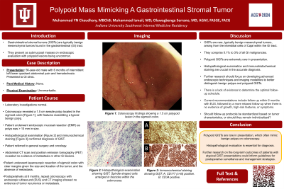
Introduction: Gastrointestinal stromal tumors (GISTs) are mesenchymal tumors that can arise anywhere in the gastrointestinal (GI) tract. They typically present as submucosal masses on endoscopic evaluation with polypoid lesions being uncommon. We present a unique case of a GIST presenting as a polypoid lesion on colonoscopy, initially being misdiagnosed as a benign polyp. Subsequent histopathological examination revealed the presence of a GIST, highlighting the importance of considering this differential diagnosis in patients with colonic polyps.
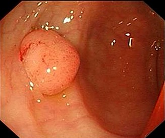
Case Description/Methods: A 56-year-old male presented to the GI clinic with a six-month history of intermittent abdominal pain and hematochezia. Physical examination was unremarkable, and laboratory investigations were within normal limits. A colonoscopy revealed a 1.5 cm polypoid lesion located in the sigmoid colon (Image 1). The lesion appeared smooth and pinkish, resembling a typical benign polyp. Histopathological examination following an endoscopic mucosal resection (EMR) demonstrated spindle-shaped cells arranged in fascicles within the submucosa, consistent with a GIST. Immunohistochemical staining was positive for CD117 (c-kit) and CD34, confirming the diagnosis. Abdominal computed tomography (CT) and positron emission tomography (PET) revealed no evidence of metastasis or other GI lesions.
Multidisciplinary management ensued, consisting of input from gastroenterologists, surgeons, and oncologists. Given the size and location of the tumor, and the absence of metastasis, the patient underwent laparoscopic resection of the sigmoid colon with clear margins. Follow-up colonoscopy and imaging studies at six months postoperatively showed no evidence of tumor recurrence or metastasis. The patient remained asymptomatic and was closely monitored in the outpatient setting.
Discussion: GISTs are rare mesenchymal tumors that arise from the interstitial cells of Cajal within the GI tract. While they typically present as submucosal masses, their presentation as polypoid lesions is unusual and can pose a diagnostic challenge.
In our case, the GIST presented as a polypoid lesion in the sigmoid colon, initially misdiagnosed as a benign polyp on colonoscopy. The accurate diagnosis was made based on histopathological examination and immunohistochemical staining, highlighting the importance of careful evaluation and consideration of GIST in the differential diagnosis of unusual endoscopic findings.

Disclosures:
Muhammad YN. Chaudhary, MBChB1, Muhammad Ismail, MD2, Oluwagbenga Serrano, MD, FACG3. P3788 - Gastrointestinal Stromal Tumor Presenting as a Submucosal Lesion Mimicking a Polyp: A Case Report, ACG 2024 Annual Scientific Meeting Abstracts. Philadelphia, PA: American College of Gastroenterology.
1Indiana University Southwest, Evansville, IN; 2Indiana University Southwest, Cedar Rapids, IA; 3Good Samaritan Hospital, Vincennes, IN
Introduction: Gastrointestinal stromal tumors (GISTs) are mesenchymal tumors that can arise anywhere in the gastrointestinal (GI) tract. They typically present as submucosal masses on endoscopic evaluation with polypoid lesions being uncommon. We present a unique case of a GIST presenting as a polypoid lesion on colonoscopy, initially being misdiagnosed as a benign polyp. Subsequent histopathological examination revealed the presence of a GIST, highlighting the importance of considering this differential diagnosis in patients with colonic polyps.
Case Description/Methods: A 56-year-old male presented to the GI clinic with a six-month history of intermittent abdominal pain and hematochezia. Physical examination was unremarkable, and laboratory investigations were within normal limits. A colonoscopy revealed a 1.5 cm polypoid lesion located in the sigmoid colon (Image 1). The lesion appeared smooth and pinkish, resembling a typical benign polyp. Histopathological examination following an endoscopic mucosal resection (EMR) demonstrated spindle-shaped cells arranged in fascicles within the submucosa, consistent with a GIST. Immunohistochemical staining was positive for CD117 (c-kit) and CD34, confirming the diagnosis. Abdominal computed tomography (CT) and positron emission tomography (PET) revealed no evidence of metastasis or other GI lesions.
Multidisciplinary management ensued, consisting of input from gastroenterologists, surgeons, and oncologists. Given the size and location of the tumor, and the absence of metastasis, the patient underwent laparoscopic resection of the sigmoid colon with clear margins. Follow-up colonoscopy and imaging studies at six months postoperatively showed no evidence of tumor recurrence or metastasis. The patient remained asymptomatic and was closely monitored in the outpatient setting.
Discussion: GISTs are rare mesenchymal tumors that arise from the interstitial cells of Cajal within the GI tract. While they typically present as submucosal masses, their presentation as polypoid lesions is unusual and can pose a diagnostic challenge.
In our case, the GIST presented as a polypoid lesion in the sigmoid colon, initially misdiagnosed as a benign polyp on colonoscopy. The accurate diagnosis was made based on histopathological examination and immunohistochemical staining, highlighting the importance of careful evaluation and consideration of GIST in the differential diagnosis of unusual endoscopic findings.

Figure: Image 1: Gastrointestinal stromal tumor (GIST) in the sigmoid colon mimicking a polyp.
Disclosures:
Muhammad Chaudhary indicated no relevant financial relationships.
Muhammad Ismail indicated no relevant financial relationships.
Oluwagbenga Serrano: MERCK – Stock-publicly held company(excluding mutual/index funds).
Muhammad YN. Chaudhary, MBChB1, Muhammad Ismail, MD2, Oluwagbenga Serrano, MD, FACG3. P3788 - Gastrointestinal Stromal Tumor Presenting as a Submucosal Lesion Mimicking a Polyp: A Case Report, ACG 2024 Annual Scientific Meeting Abstracts. Philadelphia, PA: American College of Gastroenterology.
