Tuesday Poster Session
Category: IBD
P4417 - Cytomegalovirus Isolated to a Colon Polyp in a Patient With Ulcerative Colitis on Ozanimod
Tuesday, October 29, 2024
10:30 AM - 4:00 PM ET
Location: Exhibit Hall E

Has Audio
- AM
Aun Muhammad, MD
University of Mississippi Medical Center
Jackson, MS
Presenting Author(s)
Zehra Naseem, MD1, Ahmed Nadeem, MD2, Aidan J. David, 3, Aun Muhammad, MD4, Brian M.. Fung, MD5, Stephen B. Hanauer, MD, FACG6, Joseph David, MD7
1Cleveland Clinic, Cleveland, OH; 2Cleveland Clinic Foundation, Cleveland, OH; 3Case Western Reserve University, Cleveland, OH; 4University of Mississippi Medical Center, Jackson, MS; 5Arizona Digestive Health, Mesa, AZ; 6Northwestern Medicine, Chicago, IL; 7University of Arizona College of Medicine, Phoenix, AZ
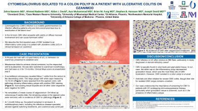
Introduction: Cytomegalovirus (CMV) infection is a notable gastrointestinal (GI) infection affecting patients who are immunocompromised due to reactivation of the latent virus. In the GI tract, CMV often presents with patchy or diffuse mucosal involvement and can cause fulminant colitis. In the following case, we describe a patient with ulcerative colitis (UC) in clinical remission on ozanimod, who was found to have CMV isolated to an inflammatory colon polyp.
Case Description/Methods: A 49-year-old man with a 2-year history of UC, in remission on ozanimod, presented to establish care. He initially failed to respond to mesalamine but achieved remission with budesonide. He subsequently was transitioned to ozanimod monotherapy and had been on it for 10 months. At presentation, his clinical Mayo score was 0.
On surveillance colonoscopy, Mayo 1 colitis was found from the rectum to the descending colon. Multiple inflamed polyps with white caps were observed in the sigmoid colon. Two of the largest polyps (13-16 mm, Figure 1A) were resected; pathology revealed inflammatory polyps with CMV colitis (Figure 1B). Biopsies of the surrounding tissue and all other colon segments were negative for CMV, with only chronic inactive colitis in the rectum and sigmoid colon. Plasma CMV PCR assay was negative. The patient completed a three-week course of valganciclovir. A follow-up colonoscopy 6 weeks later revealed similar endoscopic findings. All remaining polyps with white caps in the sigmoid colon (5-12 mm) were resected with no evidence of CMV (Figure 1C). At one-month follow-up, the patient remained in clinical remission. A multidisciplinary team advised that switching treatments would not guarantee improved safety or efficacy, and the patient elected to stay on ozanimod.
Discussion: This is the first reported case of CMV isolated to a colon polyp in a patient with UC taking ozanimod. CMV is typically found in ulcer beds and granulation tissue which explains its isolation to the ulcerated white caps of the resected polyps. Frequently, CMV in UC manifests with severe gastrointestinal symptoms, closely resembling an IBD flare. However, CMV isolated to a colon polyp is unusual and likely represents clinically silent CMV reactivation. The natural history and optimal management of isolated CMV remains unclear.

Disclosures:
Zehra Naseem, MD1, Ahmed Nadeem, MD2, Aidan J. David, 3, Aun Muhammad, MD4, Brian M.. Fung, MD5, Stephen B. Hanauer, MD, FACG6, Joseph David, MD7. P4417 - Cytomegalovirus Isolated to a Colon Polyp in a Patient With Ulcerative Colitis on Ozanimod, ACG 2024 Annual Scientific Meeting Abstracts. Philadelphia, PA: American College of Gastroenterology.
1Cleveland Clinic, Cleveland, OH; 2Cleveland Clinic Foundation, Cleveland, OH; 3Case Western Reserve University, Cleveland, OH; 4University of Mississippi Medical Center, Jackson, MS; 5Arizona Digestive Health, Mesa, AZ; 6Northwestern Medicine, Chicago, IL; 7University of Arizona College of Medicine, Phoenix, AZ
Introduction: Cytomegalovirus (CMV) infection is a notable gastrointestinal (GI) infection affecting patients who are immunocompromised due to reactivation of the latent virus. In the GI tract, CMV often presents with patchy or diffuse mucosal involvement and can cause fulminant colitis. In the following case, we describe a patient with ulcerative colitis (UC) in clinical remission on ozanimod, who was found to have CMV isolated to an inflammatory colon polyp.
Case Description/Methods: A 49-year-old man with a 2-year history of UC, in remission on ozanimod, presented to establish care. He initially failed to respond to mesalamine but achieved remission with budesonide. He subsequently was transitioned to ozanimod monotherapy and had been on it for 10 months. At presentation, his clinical Mayo score was 0.
On surveillance colonoscopy, Mayo 1 colitis was found from the rectum to the descending colon. Multiple inflamed polyps with white caps were observed in the sigmoid colon. Two of the largest polyps (13-16 mm, Figure 1A) were resected; pathology revealed inflammatory polyps with CMV colitis (Figure 1B). Biopsies of the surrounding tissue and all other colon segments were negative for CMV, with only chronic inactive colitis in the rectum and sigmoid colon. Plasma CMV PCR assay was negative. The patient completed a three-week course of valganciclovir. A follow-up colonoscopy 6 weeks later revealed similar endoscopic findings. All remaining polyps with white caps in the sigmoid colon (5-12 mm) were resected with no evidence of CMV (Figure 1C). At one-month follow-up, the patient remained in clinical remission. A multidisciplinary team advised that switching treatments would not guarantee improved safety or efficacy, and the patient elected to stay on ozanimod.
Discussion: This is the first reported case of CMV isolated to a colon polyp in a patient with UC taking ozanimod. CMV is typically found in ulcer beds and granulation tissue which explains its isolation to the ulcerated white caps of the resected polyps. Frequently, CMV in UC manifests with severe gastrointestinal symptoms, closely resembling an IBD flare. However, CMV isolated to a colon polyp is unusual and likely represents clinically silent CMV reactivation. The natural history and optimal management of isolated CMV remains unclear.

Figure: Figure 1A: Sigmoid colon inflammatory polyp 1B: Sigmoid colon polyp pathology revealing CMV inclusion bodies using immunohistochemical staining (original magnification x400)
1C: Sigmoid colon polyp with no evidence of CMV after valganciclovir treatment (original magnification x400)
1C: Sigmoid colon polyp with no evidence of CMV after valganciclovir treatment (original magnification x400)
Disclosures:
Zehra Naseem indicated no relevant financial relationships.
Ahmed Nadeem indicated no relevant financial relationships.
Aidan J. David indicated no relevant financial relationships.
Aun Muhammad indicated no relevant financial relationships.
Brian Fung indicated no relevant financial relationships.
Stephen B. Hanauer: Abbvie – Consultant. Johnson and Johnson – Consultant. Takeda – Consultant.
Joseph David indicated no relevant financial relationships.
Zehra Naseem, MD1, Ahmed Nadeem, MD2, Aidan J. David, 3, Aun Muhammad, MD4, Brian M.. Fung, MD5, Stephen B. Hanauer, MD, FACG6, Joseph David, MD7. P4417 - Cytomegalovirus Isolated to a Colon Polyp in a Patient With Ulcerative Colitis on Ozanimod, ACG 2024 Annual Scientific Meeting Abstracts. Philadelphia, PA: American College of Gastroenterology.
