Tuesday Poster Session
Category: IBD
P4420 - Don’t Claw Your Way Out: A Severe Case of Crohn’s Colitis With Bear Claw Ulcers Seen on Colonoscopy
Tuesday, October 29, 2024
10:30 AM - 4:00 PM ET
Location: Exhibit Hall E

Has Audio
- JM
Jaslyn Maurer, DO
New York-Presbyterian/Queens
Flushing, NY
Presenting Author(s)
Jaslyn Maurer, DO1, Jeffrey Dong, MD2, Mostafa Kokabee, MD3, Pamela Unger, MD3, Arun Swaminath, MD4
1New York-Presbyterian/Queens, Flushing, NY; 2Lenox Hill Hospital, Northwell Health, Long Island City, NY; 3Lenox Hill Hospital, Northwell Health, New York, NY; 4Lenox Hill Hospital/Northwell Health, New York, NY
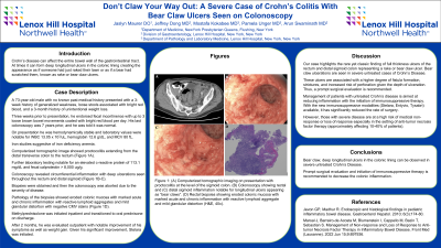
Introduction: Crohn’s disease can affect the entire bowel wall of the gastrointestinal tract. At times it can form deep longitudinal ulcers in the colonic lining creating the appearance as if someone had just raked their lawn or as if a bear had scratched them, known as rake or bear claw ulcers. We present a case of a 73-year-old male without prior diagnosis of inflammatory bowel disease was found to have bear claw ulcers on colonoscopy.
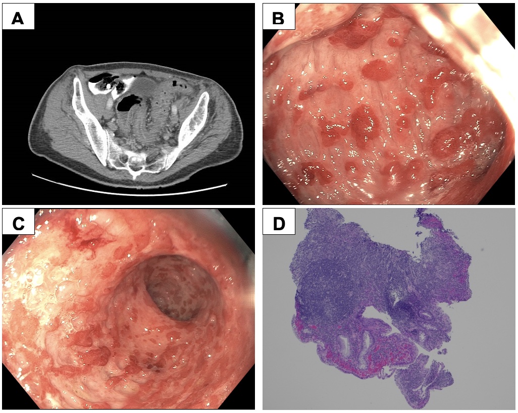
Case Description/Methods: A 73-year-old male with no known past medical history presented with a 3-week history of generalized weakness, loose stools associated with bright red blood, and a 3-month history of unintentional weight loss. Three weeks prior to presentation, he endorsed fecal incontinence with up to 3 loose brown bowel movements coated with bright red blood per day. His last colonoscopy was 7 years prior, and he was told it was normal. On presentation he was hemodynamically stable and laboratory values were notable for WBC 13.05 x 10³/uL, hemoglobin 12.6 g/dL, and MCV 80 fL. Iron studies suggestive of iron deficiency anemia. Computerized tomographic image showed proctocolitis extending from the distal transverse colon to the rectum (Figure 1A). Further laboratory testing notable for an elevated c-reactive protein of 113.1 mg/dL and fecal calprotectin > 8,000 ug/g. Colonoscopy revealed circumferential inflammation with deep ulcerations seen throughout the rectum and distal sigmoid (Figure 1B-C). Biopsies were obtained and then the colonoscopy was aborted due to the severity of disease. Pathology of the biopsies showed eroded colonic mucosa with marked acute and chronic inflammation with reactive lymphoid aggregates and mild glandular distortion with negative CMV stains (Figure 1D). Methylprednisolone was initiated inpatient and transitioned to oral prednisone on discharge. After 2 months, he was evaluated outpatient with notable improvement of his symptoms as well as weight gain. Given his significant improvement, Stelara was initiated.
Discussion: Our case highlights the rare yet classic finding of full thickness ulcers of the rectum and distal sigmoid colon representing a rake or bear claw ulcer. Bear claw ulcerations are seen in severe untreated cases of Crohn’s Disease. They are associated with a higher degree of fistula formation, strictures, and perforation given the depth of ulceration. Prompt surgical evaluation and initiation of immunosuppressive therapy with anti-TNF inhibitors is recommended for higher-risk disease and to induce and maintain remission.

Disclosures:
Jaslyn Maurer, DO1, Jeffrey Dong, MD2, Mostafa Kokabee, MD3, Pamela Unger, MD3, Arun Swaminath, MD4. P4420 - Don’t Claw Your Way Out: A Severe Case of Crohn’s Colitis With Bear Claw Ulcers Seen on Colonoscopy, ACG 2024 Annual Scientific Meeting Abstracts. Philadelphia, PA: American College of Gastroenterology.
1New York-Presbyterian/Queens, Flushing, NY; 2Lenox Hill Hospital, Northwell Health, Long Island City, NY; 3Lenox Hill Hospital, Northwell Health, New York, NY; 4Lenox Hill Hospital/Northwell Health, New York, NY
Introduction: Crohn’s disease can affect the entire bowel wall of the gastrointestinal tract. At times it can form deep longitudinal ulcers in the colonic lining creating the appearance as if someone had just raked their lawn or as if a bear had scratched them, known as rake or bear claw ulcers. We present a case of a 73-year-old male without prior diagnosis of inflammatory bowel disease was found to have bear claw ulcers on colonoscopy.
Case Description/Methods: A 73-year-old male with no known past medical history presented with a 3-week history of generalized weakness, loose stools associated with bright red blood, and a 3-month history of unintentional weight loss. Three weeks prior to presentation, he endorsed fecal incontinence with up to 3 loose brown bowel movements coated with bright red blood per day. His last colonoscopy was 7 years prior, and he was told it was normal. On presentation he was hemodynamically stable and laboratory values were notable for WBC 13.05 x 10³/uL, hemoglobin 12.6 g/dL, and MCV 80 fL. Iron studies suggestive of iron deficiency anemia. Computerized tomographic image showed proctocolitis extending from the distal transverse colon to the rectum (Figure 1A). Further laboratory testing notable for an elevated c-reactive protein of 113.1 mg/dL and fecal calprotectin > 8,000 ug/g. Colonoscopy revealed circumferential inflammation with deep ulcerations seen throughout the rectum and distal sigmoid (Figure 1B-C). Biopsies were obtained and then the colonoscopy was aborted due to the severity of disease. Pathology of the biopsies showed eroded colonic mucosa with marked acute and chronic inflammation with reactive lymphoid aggregates and mild glandular distortion with negative CMV stains (Figure 1D). Methylprednisolone was initiated inpatient and transitioned to oral prednisone on discharge. After 2 months, he was evaluated outpatient with notable improvement of his symptoms as well as weight gain. Given his significant improvement, Stelara was initiated.
Discussion: Our case highlights the rare yet classic finding of full thickness ulcers of the rectum and distal sigmoid colon representing a rake or bear claw ulcer. Bear claw ulcerations are seen in severe untreated cases of Crohn’s Disease. They are associated with a higher degree of fistula formation, strictures, and perforation given the depth of ulceration. Prompt surgical evaluation and initiation of immunosuppressive therapy with anti-TNF inhibitors is recommended for higher-risk disease and to induce and maintain remission.

Figure: Figure 1: (A) Computerized tomographic imaging on presentation with proctocolitis at the level of the sigmoid colon. (B) Colonoscopy showing rectal and (C) distal sigmoid inflammation notable for longitudinal ulcers appearing as “bear claws”. (D) Rectal biopsies showing eroded colonic mucosa with marked acute and chronic inflammation with reactive lymphoid aggregate and mild glandular distortion (H&E, 40x).
Disclosures:
Jaslyn Maurer indicated no relevant financial relationships.
Jeffrey Dong indicated no relevant financial relationships.
Mostafa Kokabee indicated no relevant financial relationships.
Pamela Unger indicated no relevant financial relationships.
Arun Swaminath: abbvie – Grant/Research Support. janssen – Grant/Research Support.
Jaslyn Maurer, DO1, Jeffrey Dong, MD2, Mostafa Kokabee, MD3, Pamela Unger, MD3, Arun Swaminath, MD4. P4420 - Don’t Claw Your Way Out: A Severe Case of Crohn’s Colitis With Bear Claw Ulcers Seen on Colonoscopy, ACG 2024 Annual Scientific Meeting Abstracts. Philadelphia, PA: American College of Gastroenterology.
