Tuesday Poster Session
Category: Interventional Endoscopy
P4531 - Use of SEMS in an Esophageal Perforation for Post Dilation in Type A Tracheoesophageal Fistula
Tuesday, October 29, 2024
10:30 AM - 4:00 PM ET
Location: Exhibit Hall E

Has Audio

Naren Nallapeta, MD
University at Buffalo
Buffalo, NY
Presenting Author(s)
Naren Nallapeta, MD1, Navpreet Rana, DO1, Clive J. Miranda, DO, MSc2, Alexander M. Carlson, DO1, Ali A. Aijaz, DO1, Nariman Hossein-Javaheri, DO1, Srushti Sahukar, MBBS3, Kevin Yang, MD1, Ramon Rivera, MD1
1University at Buffalo, Buffalo, NY; 2CHI Health Creighton University Medical Center, Omaha, NE; 3SUNY Upstate Medical University, North Billerica, MA
Introduction: VACTERL (vertebral defects, anal atresia, cardiac defects, trachea-esophageal fistula, renal anomalies and limb abnormalities) association is a multisystem congenital syndrome with incidence of 1/10,000 to 1/40,000 live births annually. Tracheoesophageal fistula (TEF) is noted in about 50-80%. Severe esophageal atresia is commonly treated with surgery, while less severe presentations are treated with serial balloon dilation via endoscopy.
Case Description/Methods: 14-month-old female with VACTERL association, imperforate anus with recto-vestibular fistula at birth status post perineal body sparing mini posterior sagittal anorectoplasty, bilateral grade 3 vesicoureteral fistula, tethered cord status post release at 4 months of age presented to the surgery clinic at the age of 13 months with concerns of intolerance to solid food. Esophagram showed moderately dilated esophagus with significant narrowing at the gastroesophageal junction (GEJ) concerning for esophageal atresia. EGD performed by the pediatric gastroenterologist showed a stenosis at the GEJ consistent with Type A TEF. This was dilated using a through the scope (TTS) balloon dilator up to 12 mm with heme effect. She was discharged post procedure.
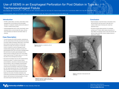
She returned to the hospital in few hours with increased fussiness, left sided neck swelling. CT neck/torso showed moderate pneumomediastinum, subcutaneous emphysema along the neck, chest and abdomen. Follow up esophagram showed persistent narrowing in the distal esophagus with extraluminal pooling of contrast just above the diaphragm consistent with an esophageal tear. Upon repeat endoscopy there was a 15mm defect along the right lateral border of the esophagus just proximal to the GEJ at 21cm. It was determined that endoscopic suturing would not be feasible due to the location of the defect and therefore a fully covered biliary self-expanding metal stent (SEMS) 10mm x 6cm was deployed under fluoroscopic and endoscopic guidance. The stent was secured using four spiral tacks and one suture.
Repeat esophagram 3 days later showed no extravasation and the diet was advanced. Stent was removed 15 days later and no evidence of defect noted on fluoroscopy then. Growth was appropriate at age 2.
Discussion: We present successful stepwise approach of an esophageal perforation post TTS balloon dilation treated with placement of a biliary SEMS in the esophagus sutured in place with spiral tacks. Off-label use of biliary stents either covered or partially covered can address TEF in children.

Disclosures:
Naren Nallapeta, MD1, Navpreet Rana, DO1, Clive J. Miranda, DO, MSc2, Alexander M. Carlson, DO1, Ali A. Aijaz, DO1, Nariman Hossein-Javaheri, DO1, Srushti Sahukar, MBBS3, Kevin Yang, MD1, Ramon Rivera, MD1. P4531 - Use of SEMS in an Esophageal Perforation for Post Dilation in Type A Tracheoesophageal Fistula, ACG 2024 Annual Scientific Meeting Abstracts. Philadelphia, PA: American College of Gastroenterology.
1University at Buffalo, Buffalo, NY; 2CHI Health Creighton University Medical Center, Omaha, NE; 3SUNY Upstate Medical University, North Billerica, MA
Introduction: VACTERL (vertebral defects, anal atresia, cardiac defects, trachea-esophageal fistula, renal anomalies and limb abnormalities) association is a multisystem congenital syndrome with incidence of 1/10,000 to 1/40,000 live births annually. Tracheoesophageal fistula (TEF) is noted in about 50-80%. Severe esophageal atresia is commonly treated with surgery, while less severe presentations are treated with serial balloon dilation via endoscopy.
Case Description/Methods: 14-month-old female with VACTERL association, imperforate anus with recto-vestibular fistula at birth status post perineal body sparing mini posterior sagittal anorectoplasty, bilateral grade 3 vesicoureteral fistula, tethered cord status post release at 4 months of age presented to the surgery clinic at the age of 13 months with concerns of intolerance to solid food. Esophagram showed moderately dilated esophagus with significant narrowing at the gastroesophageal junction (GEJ) concerning for esophageal atresia. EGD performed by the pediatric gastroenterologist showed a stenosis at the GEJ consistent with Type A TEF. This was dilated using a through the scope (TTS) balloon dilator up to 12 mm with heme effect. She was discharged post procedure.
She returned to the hospital in few hours with increased fussiness, left sided neck swelling. CT neck/torso showed moderate pneumomediastinum, subcutaneous emphysema along the neck, chest and abdomen. Follow up esophagram showed persistent narrowing in the distal esophagus with extraluminal pooling of contrast just above the diaphragm consistent with an esophageal tear. Upon repeat endoscopy there was a 15mm defect along the right lateral border of the esophagus just proximal to the GEJ at 21cm. It was determined that endoscopic suturing would not be feasible due to the location of the defect and therefore a fully covered biliary self-expanding metal stent (SEMS) 10mm x 6cm was deployed under fluoroscopic and endoscopic guidance. The stent was secured using four spiral tacks and one suture.
Repeat esophagram 3 days later showed no extravasation and the diet was advanced. Stent was removed 15 days later and no evidence of defect noted on fluoroscopy then. Growth was appropriate at age 2.
Discussion: We present successful stepwise approach of an esophageal perforation post TTS balloon dilation treated with placement of a biliary SEMS in the esophagus sutured in place with spiral tacks. Off-label use of biliary stents either covered or partially covered can address TEF in children.

Figure: Esophageal perforation post dilation treated with SEMS
Disclosures:
Naren Nallapeta indicated no relevant financial relationships.
Navpreet Rana indicated no relevant financial relationships.
Clive Miranda indicated no relevant financial relationships.
Alexander Carlson indicated no relevant financial relationships.
Ali Aijaz indicated no relevant financial relationships.
Nariman Hossein-Javaheri indicated no relevant financial relationships.
Srushti Sahukar indicated no relevant financial relationships.
Kevin Yang indicated no relevant financial relationships.
Ramon Rivera indicated no relevant financial relationships.
Naren Nallapeta, MD1, Navpreet Rana, DO1, Clive J. Miranda, DO, MSc2, Alexander M. Carlson, DO1, Ali A. Aijaz, DO1, Nariman Hossein-Javaheri, DO1, Srushti Sahukar, MBBS3, Kevin Yang, MD1, Ramon Rivera, MD1. P4531 - Use of SEMS in an Esophageal Perforation for Post Dilation in Type A Tracheoesophageal Fistula, ACG 2024 Annual Scientific Meeting Abstracts. Philadelphia, PA: American College of Gastroenterology.
