Tuesday Poster Session
Category: Interventional Endoscopy
P4540 - Catching a Wolf in Sheep’s Clothing: Successful Endoscopic Mucosal Resection of a Large Duodenal Pyloric Gland Adenoma
Tuesday, October 29, 2024
10:30 AM - 4:00 PM ET
Location: Exhibit Hall E

Has Audio
- SH
Spencer Harris, MD, PhD
Virginia Commonwealth University Health System
Richmond, VA
Presenting Author(s)
Spencer Harris, MD, PhD1, Dan McEntire, MD2, Ravi Vachhani, MD1
1Virginia Commonwealth University Health System, Richmond, VA; 2Virginia Commonwealth University Medical Center, Richmond, VA
Introduction: Pyloric gland adenomas are rare premalignant masses that arise from gastric mucosa. Most frequently they are identified in the stomach, however extragastric pyloric gland adenomas have been reported in the literature to occur in the esophagus, duodenum, and even bile ducts. Here we describe a case of a large pyloric gland adenoma identified in the duodenal bulb that was effectively treated through endoscopic removal.
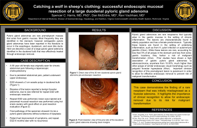
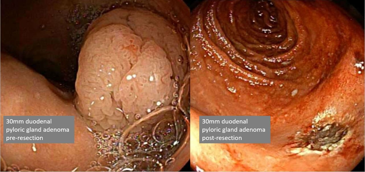
Case Description/Methods: The patient was originally seen for laparoscopic cholecystectomy due to abdominal pain six months prior to being seen by our service. Due to persistent abdominal pain, the patient subsequently underwent an upper endoscopy. This showed a large 3 cm sessile polyp in the duodenal bulb (Figure), with initial pathology results of biopsies showing benign foveolar adenoma. However, given the size, the patient was subsequently referred for repeat EGD and underwent endoscopic resection of the polyp. The lesion was raised via injection and then piecemeal mucosal resection was performed using hot snare cautery resulting in complete resection (Figure). The final pathology of the specimen confirmed it to be a pyloric gland adenoma without dysplasia.
Discussion: Pyloric gland adenomas are rare neoplasms that typically arise in the gastric mucosa in the setting of chronic inflammation. The lesions are characteristically found in elderly population and has a female predominance. Typically these lesions are found in the setting of underlying inflammation, such as from H. pylori infection or autoimmune gastritis. While these lesions are rare, accounting for less than 5% of all polyps in the stomach and less from extra-gastric locations, they harbor a risk of malignant transformation. Prior studies have shown a high association of gastric pyloric gland adenomas to adenocarcinoma, anywhere from 10-30%, much higher than other polyps of similar endoscopic visual morphology such as polypoid foveolar hyperplasia. Maintaining a clinical suspicion for this premalignant lesion is important in order to identify and remove to prevent risk of malignant transformation.

Disclosures:
Spencer Harris, MD, PhD1, Dan McEntire, MD2, Ravi Vachhani, MD1. P4540 - Catching a Wolf in Sheep’s Clothing: Successful Endoscopic Mucosal Resection of a Large Duodenal Pyloric Gland Adenoma, ACG 2024 Annual Scientific Meeting Abstracts. Philadelphia, PA: American College of Gastroenterology.
1Virginia Commonwealth University Health System, Richmond, VA; 2Virginia Commonwealth University Medical Center, Richmond, VA
Introduction: Pyloric gland adenomas are rare premalignant masses that arise from gastric mucosa. Most frequently they are identified in the stomach, however extragastric pyloric gland adenomas have been reported in the literature to occur in the esophagus, duodenum, and even bile ducts. Here we describe a case of a large pyloric gland adenoma identified in the duodenal bulb that was effectively treated through endoscopic removal.
Case Description/Methods: The patient was originally seen for laparoscopic cholecystectomy due to abdominal pain six months prior to being seen by our service. Due to persistent abdominal pain, the patient subsequently underwent an upper endoscopy. This showed a large 3 cm sessile polyp in the duodenal bulb (Figure), with initial pathology results of biopsies showing benign foveolar adenoma. However, given the size, the patient was subsequently referred for repeat EGD and underwent endoscopic resection of the polyp. The lesion was raised via injection and then piecemeal mucosal resection was performed using hot snare cautery resulting in complete resection (Figure). The final pathology of the specimen confirmed it to be a pyloric gland adenoma without dysplasia.
Discussion: Pyloric gland adenomas are rare neoplasms that typically arise in the gastric mucosa in the setting of chronic inflammation. The lesions are characteristically found in elderly population and has a female predominance. Typically these lesions are found in the setting of underlying inflammation, such as from H. pylori infection or autoimmune gastritis. While these lesions are rare, accounting for less than 5% of all polyps in the stomach and less from extra-gastric locations, they harbor a risk of malignant transformation. Prior studies have shown a high association of gastric pyloric gland adenomas to adenocarcinoma, anywhere from 10-30%, much higher than other polyps of similar endoscopic visual morphology such as polypoid foveolar hyperplasia. Maintaining a clinical suspicion for this premalignant lesion is important in order to identify and remove to prevent risk of malignant transformation.

Figure: Endoscopic images of the large 3 cm duodenal pyloric gland adenoma before and after successful endoscopic mucosal resection.
Disclosures:
Spencer Harris indicated no relevant financial relationships.
Dan McEntire indicated no relevant financial relationships.
Ravi Vachhani indicated no relevant financial relationships.
Spencer Harris, MD, PhD1, Dan McEntire, MD2, Ravi Vachhani, MD1. P4540 - Catching a Wolf in Sheep’s Clothing: Successful Endoscopic Mucosal Resection of a Large Duodenal Pyloric Gland Adenoma, ACG 2024 Annual Scientific Meeting Abstracts. Philadelphia, PA: American College of Gastroenterology.
