Tuesday Poster Session
Category: Interventional Endoscopy
P4560 - Development of a Large Tracheoesophageal Fistula After Thoracic Endovascular Aortic Repair (TEVAR)
Tuesday, October 29, 2024
10:30 AM - 4:00 PM ET
Location: Exhibit Hall E

Has Audio

Julianna Tantum, DO
Lankenau Medical Center
Wynnewood, PA
Presenting Author(s)
Julianna Tantum, DO, Stefanie Gallagher, DO, Lauren Davis, DO, Erin Hollis, DO, Babek Etemad, MD
Lankenau Medical Center, Wynnewood, PA
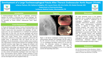
Introduction: A tracheoesophageal fistula (TEF) is a pathologic connection between the trachea or bronchial tree and the esophagus. We present a case of a patient diagnosed with a TEF, hypothesized to be secondary to recent thoracic endovascular aortic repair (TEVAR).
Case Description/Methods: This is a 79-year-old female with history of venous thromboembolism with IVC filter (on rivaroxaban) and thoracic aortic aneurysm status post TEVAR six weeks prior who presented with dysphagia.
Of note, one month prior to this presentation, she was hospitalized for an endoleak and underwent TEVAR repair. At that time, she had new dysphagia with barium swallow study showing narrowing of the midesophagus, likely mass effect from known thoracic aortic aneurysm, with extrinsic compression confirmed on endoscopic evaluation performed by the surgical team. A gastrostomy tube was placed, and the patient was discharged.
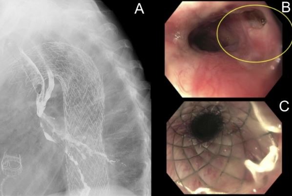
On this presentation, a barium swallow study revealed new, large fistulous communication between the esophagus and left mainstem bronchus origin (Figure 1A). Imaging also showed a left sided pneumonia, treated with antibiotics. She was evaluated by interventional pulmonology and gastroenterology and underwent tandem stent placement with tracheal stent placement with bronchoscopy followed by endoscopic placement of a 14 mm x 12 cm fully covered self-expanding esophageal metal stent (Figure 1B &1C). Post-procedure barium swallow showed resolution of prior fistula. She continued with tube feeds and tolerated a full liquid diet and was discharged with close outpatient follow up
Discussion: TEF most commonly occurs in the pediatric population due to congenital abnormalities but also, can be acquired in adulthood due to esophageal malignancy, cancer therapy, prolonged endotracheal intubation, traumatic airway injury, granulomatous mediastinal infection, or ingestion of foreign bodies or corrosive products. Case reports have noted development of TEF in association with aortic aneurysms, but no data exist for their development in association with TEVAR. Patients with TEF typically present with dysphagia, coughing, pneumonia, and chest pain. Diagnosis may include thoracic imaging with barium esophagram and upper endoscopy with flexible bronchoscopy. Treatment options include tracheal and esophageal stenting or surgery.
Kim HS, Khemasuwan D,Diaz-Mendoza J, Mehta AC.Management of tracheo-oesophageal fistula in adults.Eur Respir Rev.2020 Nov 5;29(158).

Disclosures:
Julianna Tantum, DO, Stefanie Gallagher, DO, Lauren Davis, DO, Erin Hollis, DO, Babek Etemad, MD. P4560 - Development of a Large Tracheoesophageal Fistula After Thoracic Endovascular Aortic Repair (TEVAR), ACG 2024 Annual Scientific Meeting Abstracts. Philadelphia, PA: American College of Gastroenterology.
Lankenau Medical Center, Wynnewood, PA
Introduction: A tracheoesophageal fistula (TEF) is a pathologic connection between the trachea or bronchial tree and the esophagus. We present a case of a patient diagnosed with a TEF, hypothesized to be secondary to recent thoracic endovascular aortic repair (TEVAR).
Case Description/Methods: This is a 79-year-old female with history of venous thromboembolism with IVC filter (on rivaroxaban) and thoracic aortic aneurysm status post TEVAR six weeks prior who presented with dysphagia.
Of note, one month prior to this presentation, she was hospitalized for an endoleak and underwent TEVAR repair. At that time, she had new dysphagia with barium swallow study showing narrowing of the midesophagus, likely mass effect from known thoracic aortic aneurysm, with extrinsic compression confirmed on endoscopic evaluation performed by the surgical team. A gastrostomy tube was placed, and the patient was discharged.
On this presentation, a barium swallow study revealed new, large fistulous communication between the esophagus and left mainstem bronchus origin (Figure 1A). Imaging also showed a left sided pneumonia, treated with antibiotics. She was evaluated by interventional pulmonology and gastroenterology and underwent tandem stent placement with tracheal stent placement with bronchoscopy followed by endoscopic placement of a 14 mm x 12 cm fully covered self-expanding esophageal metal stent (Figure 1B &1C). Post-procedure barium swallow showed resolution of prior fistula. She continued with tube feeds and tolerated a full liquid diet and was discharged with close outpatient follow up
Discussion: TEF most commonly occurs in the pediatric population due to congenital abnormalities but also, can be acquired in adulthood due to esophageal malignancy, cancer therapy, prolonged endotracheal intubation, traumatic airway injury, granulomatous mediastinal infection, or ingestion of foreign bodies or corrosive products. Case reports have noted development of TEF in association with aortic aneurysms, but no data exist for their development in association with TEVAR. Patients with TEF typically present with dysphagia, coughing, pneumonia, and chest pain. Diagnosis may include thoracic imaging with barium esophagram and upper endoscopy with flexible bronchoscopy. Treatment options include tracheal and esophageal stenting or surgery.
Kim HS, Khemasuwan D,Diaz-Mendoza J, Mehta AC.Management of tracheo-oesophageal fistula in adults.Eur Respir Rev.2020 Nov 5;29(158).

Figure:
Figure 1: (A) Barium esophagram showing large fistulous communication between the esophagus and the origin of the left mainstem bronchus; (B) endoscopically visualized ~1.5 cm esophageal wall defect noted at 27 cm from the incisors. No endoscopic evidence of malignancy with remaining esophagus unremarkable; (C) view of endoscopically-placed esophageal 14 mm x 12 cm long fully covered self-expanding metal stent.
Figure 1: (A) Barium esophagram showing large fistulous communication between the esophagus and the origin of the left mainstem bronchus; (B) endoscopically visualized ~1.5 cm esophageal wall defect noted at 27 cm from the incisors. No endoscopic evidence of malignancy with remaining esophagus unremarkable; (C) view of endoscopically-placed esophageal 14 mm x 12 cm long fully covered self-expanding metal stent.
Disclosures:
Julianna Tantum indicated no relevant financial relationships.
Stefanie Gallagher indicated no relevant financial relationships.
Lauren Davis indicated no relevant financial relationships.
Erin Hollis indicated no relevant financial relationships.
Babek Etemad indicated no relevant financial relationships.
Julianna Tantum, DO, Stefanie Gallagher, DO, Lauren Davis, DO, Erin Hollis, DO, Babek Etemad, MD. P4560 - Development of a Large Tracheoesophageal Fistula After Thoracic Endovascular Aortic Repair (TEVAR), ACG 2024 Annual Scientific Meeting Abstracts. Philadelphia, PA: American College of Gastroenterology.
