Tuesday Poster Session
Category: Liver
P4738 - Evidence of Iron Overload After COVID-19 Vaccinations: The Li(Fe) of Identical Twins
Tuesday, October 29, 2024
10:30 AM - 4:00 PM ET
Location: Exhibit Hall E

Has Audio

Soyoun J. Pak, MD
Brooke Army Medical Center
Converse, TX
Presenting Author(s)
Soyoun Pak, MD1, Jessica Basso, MD2, Ryan Smith, DO2, Benjamin Ramos, MD2, Cassandra L. Craig, MD3
1Brooke Army Medical Center, Converse, TX; 2Brooke Army Medical Center, San Antonio, TX; 3Wilford Hall Ambulatory Surgical Center, San Antonio, TX
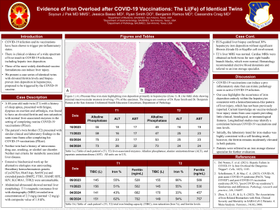
Introduction: COVID-19 infection and vaccinations have been shown to trigger pro-inflammatory states. There is clinical evidence of liver insult in COVID-19 infections, including hepatic iron deposition. Also, all three of the most widely distributed vaccine formulations can induce liver injury. This is a case series of identical twins with elevated ferritin levels and biopsy-proven iron deposition in hepatocytes, proposed to be triggered by the COVID-19 vaccine.
Case Description/Methods: A 48-year-old male twin (T1), presenting with fatigue, dyspnea on exertion and arthralgia, was found to have an elevated ferritin and iron saturation with normal liver-associated enzymes after routine COVID-19 vaccinations (Pfizer). The patient’s twin brother (T2) presented with similar clinical and laboratory findings in the same time frame after completing COVID-19 vaccinations (Pfizer). Neither twin had a history of intravenous drug-use, smoking, or alcohol use disorder. Neither met criteria for metabolic associated liver disease. Extensive biochemical work up for alternative etiologies was unrevealing. Hereditary hemochromatosis genetic panels (Cys282Tyr, His63Asp, Ser65Cys, BMP2, FTH1, HAMP, HFE, HJV, SLC40A1, TFR2) were unremarkable. Abdominal ultrasound showed normal liver morphology. T1’s magnetic resonance liver with elastography (MRE) revealed iron concentration of 2.3 mg/g (normal < 2 mg/g) with composite value of 1.8 kPa, and liver biopsy confirmed 30% hepatocyte iron deposition without significant fibrosis or Kupffer cell involvement. T2’s liver MRE was normal. Cardiac MRIs were obtained for new right bundle branch blocks and were normal. The patients were co-managed with hematology, who recommended considering elective blood donations and referral to an iron storage disease specialist.
Discussion: COVID-19 vaccinations can induce a pro-inflammatory state that can mimic pathology seen in active COVID-19 infection. Current knowledge on vaccine-induced-liver injury stems from case reports - providing little clinical, histological, or immunological data. Our case highlights iron deposition entirely within the hepatocytes consistent with a hemochromatosis-like pattern of liver injury, which has not been previously reported. The patients were without concomitant chronic liver disease and had no other risk factors for chronic liver disease. Although initially the laboratory trend for iron studies was highly consistent with a self-limiting insult, the ferritin remains moderately elevated in both patients.

Note: The table for this abstract can be viewed in the ePoster Gallery section of the ACG 2024 ePoster Site or in The American Journal of Gastroenterology's abstract supplement issue, both of which will be available starting October 27, 2024.
Disclosures:
Soyoun Pak, MD1, Jessica Basso, MD2, Ryan Smith, DO2, Benjamin Ramos, MD2, Cassandra L. Craig, MD3. P4738 - Evidence of Iron Overload After COVID-19 Vaccinations: The Li(Fe) of Identical Twins, ACG 2024 Annual Scientific Meeting Abstracts. Philadelphia, PA: American College of Gastroenterology.
1Brooke Army Medical Center, Converse, TX; 2Brooke Army Medical Center, San Antonio, TX; 3Wilford Hall Ambulatory Surgical Center, San Antonio, TX
Introduction: COVID-19 infection and vaccinations have been shown to trigger pro-inflammatory states. There is clinical evidence of liver insult in COVID-19 infections, including hepatic iron deposition. Also, all three of the most widely distributed vaccine formulations can induce liver injury. This is a case series of identical twins with elevated ferritin levels and biopsy-proven iron deposition in hepatocytes, proposed to be triggered by the COVID-19 vaccine.
Case Description/Methods: A 48-year-old male twin (T1), presenting with fatigue, dyspnea on exertion and arthralgia, was found to have an elevated ferritin and iron saturation with normal liver-associated enzymes after routine COVID-19 vaccinations (Pfizer). The patient’s twin brother (T2) presented with similar clinical and laboratory findings in the same time frame after completing COVID-19 vaccinations (Pfizer). Neither twin had a history of intravenous drug-use, smoking, or alcohol use disorder. Neither met criteria for metabolic associated liver disease. Extensive biochemical work up for alternative etiologies was unrevealing. Hereditary hemochromatosis genetic panels (Cys282Tyr, His63Asp, Ser65Cys, BMP2, FTH1, HAMP, HFE, HJV, SLC40A1, TFR2) were unremarkable. Abdominal ultrasound showed normal liver morphology. T1’s magnetic resonance liver with elastography (MRE) revealed iron concentration of 2.3 mg/g (normal < 2 mg/g) with composite value of 1.8 kPa, and liver biopsy confirmed 30% hepatocyte iron deposition without significant fibrosis or Kupffer cell involvement. T2’s liver MRE was normal. Cardiac MRIs were obtained for new right bundle branch blocks and were normal. The patients were co-managed with hematology, who recommended considering elective blood donations and referral to an iron storage disease specialist.
Discussion: COVID-19 vaccinations can induce a pro-inflammatory state that can mimic pathology seen in active COVID-19 infection. Current knowledge on vaccine-induced-liver injury stems from case reports - providing little clinical, histological, or immunological data. Our case highlights iron deposition entirely within the hepatocytes consistent with a hemochromatosis-like pattern of liver injury, which has not been previously reported. The patients were without concomitant chronic liver disease and had no other risk factors for chronic liver disease. Although initially the laboratory trend for iron studies was highly consistent with a self-limiting insult, the ferritin remains moderately elevated in both patients.

Figure: A.) Prussian blue iron stain highlighting iron deposition primarily in hepatocytes (Zone 1). B.) An H&E slide showing minimal macrovesicular steatosis, involving <5% of the specimen. The images are courtesy of Dr. Ryan Smith at the San Antonio Uniformed Health Education Consortium, Department of Pathology.
Note: The table for this abstract can be viewed in the ePoster Gallery section of the ACG 2024 ePoster Site or in The American Journal of Gastroenterology's abstract supplement issue, both of which will be available starting October 27, 2024.
Disclosures:
Soyoun Pak indicated no relevant financial relationships.
Jessica Basso indicated no relevant financial relationships.
Ryan Smith indicated no relevant financial relationships.
Benjamin Ramos indicated no relevant financial relationships.
Cassandra Craig indicated no relevant financial relationships.
Soyoun Pak, MD1, Jessica Basso, MD2, Ryan Smith, DO2, Benjamin Ramos, MD2, Cassandra L. Craig, MD3. P4738 - Evidence of Iron Overload After COVID-19 Vaccinations: The Li(Fe) of Identical Twins, ACG 2024 Annual Scientific Meeting Abstracts. Philadelphia, PA: American College of Gastroenterology.
