Tuesday Poster Session
Category: Liver
P4777 - Steatotic Mass Secondary to Heterozygous Hemochromatosis, A Rare Case of Biliary Obstruction
Tuesday, October 29, 2024
10:30 AM - 4:00 PM ET
Location: Exhibit Hall E

Has Audio

Saddam Zaidi, MD
Bridgeport Hospital
Trumbull, CT
Presenting Author(s)
Saddam Zaidi, MD1, Cheng-Hung Tai, MD2, Joanna Gibson, MD, PhD3, Caroline Loeser, MD2
1Bridgeport Hospital, Trumbull, CT; 2Yale New Haven Health, Bridgeport Hospital, Bridgeport, CT; 3Yale University School of Medicine, New Haven, CT
Introduction: Heterozygous Hemochromatosis (HH) is an autosomal recessive genetic disorder causing increased iron absorption and overload. Carriers of the C282Y HFE gene mutation are at higher risk for nonalcoholic fatty liver disease (NAFLD). Studies highlight a significant link between steatosis and fibrosis in HH. Here we present a 31-year-old male with unusual biliary obstruction secondary to steatotic infiltration.
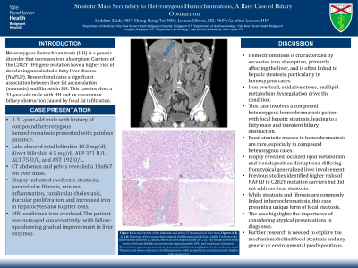
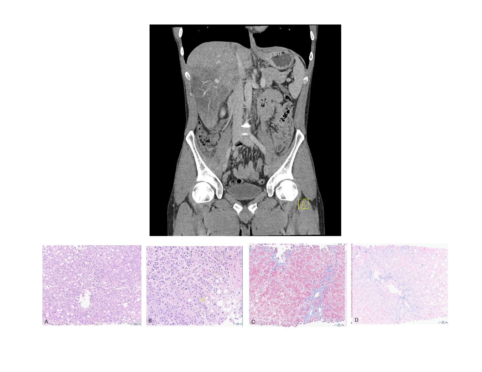
Case Description/Methods: A 31-year-old male with history of compound heterozygous hemochromatosis presented with painless jaundice. Labs showed total bilirubin 10.3 mg/dl, direct bilirubin 4.5 mg/dl, ALP 371 U/L, ALT 75 U/L, and AST 192 U/L. CT abdomen and pelvis revealed a 14x8x7 cm mass. Biopsy indicated moderate steatosis, paracellular fibrosis, minimal inflammation, canalicular cholestasis, ductular proliferation, and increased iron in hepatocytes and Kupffer cells. MRI confirmed iron overload. The patient was managed conservatively, with follow-ups showing gradual improvement in liver enzymes.
Discussion: Hemochromatosis is characterized by excessive iron absorption and deposition in organs, mainly the liver. It is associated with hepatic steatosis, particularly in homozygous forms, due to the interplay between iron overload, oxidative stress, and lipid metabolism dysregulation. This case is unique as it involves a compound heterozygous hemochromatosis patient with focal hepatic steatosis, resulting in a fatty mass causing transient biliary obstruction.
Limited data exist on focal steatotic masses in hemochromatosis, especially in compound heterozygous cases, adding complexity to disease pathology understanding. The biopsy findings suggest a localized disruption in lipid metabolism and iron deposition, diverging from the usual generalized hepatic involvement. A 2003 study indicated that C282Y mutation carriers of the HFE gene have a higher risk of NAFLD but did not address focal steatosis. Another study showed a significant link between steatosis and fibrosis in hemochromatosis, suggesting a generalized liver involvement. In this case, focal steatosis deviates from these findings, suggesting a unique manifestation.
This highlights the potential for atypical presentations and the importance of considering focal hepatic conditions in differential diagnoses. Further research is needed to explore mechanisms driving focal steatosis in hemochromatosis and whether genetic or environmental factors predispose patients to this rare presentation.

Disclosures:
Saddam Zaidi, MD1, Cheng-Hung Tai, MD2, Joanna Gibson, MD, PhD3, Caroline Loeser, MD2. P4777 - Steatotic Mass Secondary to Heterozygous Hemochromatosis, A Rare Case of Biliary Obstruction, ACG 2024 Annual Scientific Meeting Abstracts. Philadelphia, PA: American College of Gastroenterology.
1Bridgeport Hospital, Trumbull, CT; 2Yale New Haven Health, Bridgeport Hospital, Bridgeport, CT; 3Yale University School of Medicine, New Haven, CT
Introduction: Heterozygous Hemochromatosis (HH) is an autosomal recessive genetic disorder causing increased iron absorption and overload. Carriers of the C282Y HFE gene mutation are at higher risk for nonalcoholic fatty liver disease (NAFLD). Studies highlight a significant link between steatosis and fibrosis in HH. Here we present a 31-year-old male with unusual biliary obstruction secondary to steatotic infiltration.
Case Description/Methods: A 31-year-old male with history of compound heterozygous hemochromatosis presented with painless jaundice. Labs showed total bilirubin 10.3 mg/dl, direct bilirubin 4.5 mg/dl, ALP 371 U/L, ALT 75 U/L, and AST 192 U/L. CT abdomen and pelvis revealed a 14x8x7 cm mass. Biopsy indicated moderate steatosis, paracellular fibrosis, minimal inflammation, canalicular cholestasis, ductular proliferation, and increased iron in hepatocytes and Kupffer cells. MRI confirmed iron overload. The patient was managed conservatively, with follow-ups showing gradual improvement in liver enzymes.
Discussion: Hemochromatosis is characterized by excessive iron absorption and deposition in organs, mainly the liver. It is associated with hepatic steatosis, particularly in homozygous forms, due to the interplay between iron overload, oxidative stress, and lipid metabolism dysregulation. This case is unique as it involves a compound heterozygous hemochromatosis patient with focal hepatic steatosis, resulting in a fatty mass causing transient biliary obstruction.
Limited data exist on focal steatotic masses in hemochromatosis, especially in compound heterozygous cases, adding complexity to disease pathology understanding. The biopsy findings suggest a localized disruption in lipid metabolism and iron deposition, diverging from the usual generalized hepatic involvement. A 2003 study indicated that C282Y mutation carriers of the HFE gene have a higher risk of NAFLD but did not address focal steatosis. Another study showed a significant link between steatosis and fibrosis in hemochromatosis, suggesting a generalized liver involvement. In this case, focal steatosis deviates from these findings, suggesting a unique manifestation.
This highlights the potential for atypical presentations and the importance of considering focal hepatic conditions in differential diagnoses. Further research is needed to explore mechanisms driving focal steatosis in hemochromatosis and whether genetic or environmental factors predispose patients to this rare presentation.

Figure: Figure 1: Coronal section of the Abdomen and pelvis MRI showing focal liver mass.
Figures A, B, C, & D: Histology of liver parenchyma stained with Hematoxylin & Eosin (A&B), Trichrome (B) and Prussian blue iron (C) stains, shown at 200x magnification (A, C, D). The lobular parenchyma shows mild centrilobular macrovesicular (approximately 20%) and canalicular cholestasis. There is heterogenous pericentral and perisinusoidal fibrosis highlighted by the trichrome stain. The iron stain shows siderosis in both hepatocytes (with periportal accentuation) and in Kupffer cells (overall 2+).
Figures A, B, C, & D: Histology of liver parenchyma stained with Hematoxylin & Eosin (A&B), Trichrome (B) and Prussian blue iron (C) stains, shown at 200x magnification (A, C, D). The lobular parenchyma shows mild centrilobular macrovesicular (approximately 20%) and canalicular cholestasis. There is heterogenous pericentral and perisinusoidal fibrosis highlighted by the trichrome stain. The iron stain shows siderosis in both hepatocytes (with periportal accentuation) and in Kupffer cells (overall 2+).
Disclosures:
Saddam Zaidi indicated no relevant financial relationships.
Cheng-Hung Tai indicated no relevant financial relationships.
Joanna Gibson indicated no relevant financial relationships.
Caroline Loeser indicated no relevant financial relationships.
Saddam Zaidi, MD1, Cheng-Hung Tai, MD2, Joanna Gibson, MD, PhD3, Caroline Loeser, MD2. P4777 - Steatotic Mass Secondary to Heterozygous Hemochromatosis, A Rare Case of Biliary Obstruction, ACG 2024 Annual Scientific Meeting Abstracts. Philadelphia, PA: American College of Gastroenterology.
