Tuesday Poster Session
Category: Liver
P4840 - Hepatic Lymphangiomatosis: A Case Report
Tuesday, October 29, 2024
10:30 AM - 4:00 PM ET
Location: Exhibit Hall E

Has Audio

Muhammad YN Chaudhary, MBChB
Indiana University Southwest
Evansville, IN
Presenting Author(s)
Muhammad YN. Chaudhary, MBChB1, Muhammad Ismail, MD2, Oluwagbenga Serrano, MD, FACG3
1Indiana University Southwest, Evansville, IN; 2Indiana University Southwest, Cedar Rapids, IA; 3Good Samaritan Hospital, Vincennes, IN
Introduction: Hepatic lymphangiomatosis (HL) is an exceedingly rare condition characterized by the cystic dilatation of lymphatic vessels within the liver. Its clinical presentation is often nonspecific, leading to diagnostic challenges. This report highlights a novel case of HL, aiming to enhance awareness and understanding of this rare entity among physicians.
Case Description/Methods: A 34-year-old female presented with intermittent right upper quadrant abdominal pain and occasional episodes of jaundice over the past six months. Physical examination revealed mild hepatomegaly without tenderness. Liver function tests showed mildly elevated alkaline phosphatase (220 U/L) and gamma-glutamyl transferase (150 U/L) levels.
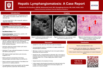
Abdominal ultrasound revealed multiple cystic lesions throughout the liver. A subsequent computed tomography (CT) scan confirmed the presence of numerous well-defined, fluid-filled cysts (Image 1). Given the unclear nature of the lesions, an MRI was performed, which demonstrated hyperintense cystic structures on T2-weighted images, suggestive of lymphangiomatosis.
To confirm the diagnosis, a percutaneous liver biopsy was conducted. Histological examination revealed cystic dilatation of lymphatic vessels with no evidence of malignancy, consistent with HL. Due to the patient's persistent symptoms and the potential for complications, surgical resection of the affected liver segments was performed. The postoperative course was uneventful, and the patient reported significant symptomatic relief.
Discussion: This case of HL is novel due to its rarity and the diagnostic challenges it presented. HL can mimic other cystic liver diseases, such as polycystic liver disease and cystic echinococcosis, leading to potential misdiagnosis. Accurate diagnosis typically requires a combination of imaging modalities and histopathological confirmation.
MRI plays a pivotal role in the non-invasive diagnosis of HL, providing superior soft tissue contrast that helps differentiate lymphatic cysts from other hepatic lesions. In this case, MRI findings were crucial in guiding the biopsy, which confirmed the diagnosis.
This report underscores the importance of considering HL in patients presenting with multiple hepatic cysts and nonspecific symptoms. Increased awareness and recognition of this condition can facilitate early diagnosis and appropriate management, thereby improving patient outcomes.

Disclosures:
Muhammad YN. Chaudhary, MBChB1, Muhammad Ismail, MD2, Oluwagbenga Serrano, MD, FACG3. P4840 - Hepatic Lymphangiomatosis: A Case Report, ACG 2024 Annual Scientific Meeting Abstracts. Philadelphia, PA: American College of Gastroenterology.
1Indiana University Southwest, Evansville, IN; 2Indiana University Southwest, Cedar Rapids, IA; 3Good Samaritan Hospital, Vincennes, IN
Introduction: Hepatic lymphangiomatosis (HL) is an exceedingly rare condition characterized by the cystic dilatation of lymphatic vessels within the liver. Its clinical presentation is often nonspecific, leading to diagnostic challenges. This report highlights a novel case of HL, aiming to enhance awareness and understanding of this rare entity among physicians.
Case Description/Methods: A 34-year-old female presented with intermittent right upper quadrant abdominal pain and occasional episodes of jaundice over the past six months. Physical examination revealed mild hepatomegaly without tenderness. Liver function tests showed mildly elevated alkaline phosphatase (220 U/L) and gamma-glutamyl transferase (150 U/L) levels.
Abdominal ultrasound revealed multiple cystic lesions throughout the liver. A subsequent computed tomography (CT) scan confirmed the presence of numerous well-defined, fluid-filled cysts (Image 1). Given the unclear nature of the lesions, an MRI was performed, which demonstrated hyperintense cystic structures on T2-weighted images, suggestive of lymphangiomatosis.
To confirm the diagnosis, a percutaneous liver biopsy was conducted. Histological examination revealed cystic dilatation of lymphatic vessels with no evidence of malignancy, consistent with HL. Due to the patient's persistent symptoms and the potential for complications, surgical resection of the affected liver segments was performed. The postoperative course was uneventful, and the patient reported significant symptomatic relief.
Discussion: This case of HL is novel due to its rarity and the diagnostic challenges it presented. HL can mimic other cystic liver diseases, such as polycystic liver disease and cystic echinococcosis, leading to potential misdiagnosis. Accurate diagnosis typically requires a combination of imaging modalities and histopathological confirmation.
MRI plays a pivotal role in the non-invasive diagnosis of HL, providing superior soft tissue contrast that helps differentiate lymphatic cysts from other hepatic lesions. In this case, MRI findings were crucial in guiding the biopsy, which confirmed the diagnosis.
This report underscores the importance of considering HL in patients presenting with multiple hepatic cysts and nonspecific symptoms. Increased awareness and recognition of this condition can facilitate early diagnosis and appropriate management, thereby improving patient outcomes.

Figure: Image 1: CT scan showing cystic lesions in the liver.
Disclosures:
Muhammad Chaudhary indicated no relevant financial relationships.
Muhammad Ismail indicated no relevant financial relationships.
Oluwagbenga Serrano: MERCK – Stock-publicly held company(excluding mutual/index funds).
Muhammad YN. Chaudhary, MBChB1, Muhammad Ismail, MD2, Oluwagbenga Serrano, MD, FACG3. P4840 - Hepatic Lymphangiomatosis: A Case Report, ACG 2024 Annual Scientific Meeting Abstracts. Philadelphia, PA: American College of Gastroenterology.
