Tuesday Poster Session
Category: Liver
P4869 - Case Study: Community-Acquired Klebsiella Liver Abscess in the Absence of Diabetes Mellitus and Hepatobiliary Disease
Tuesday, October 29, 2024
10:30 AM - 4:00 PM ET
Location: Exhibit Hall E

Has Audio

Jalal Samhoun, MD
Florida Atlantic University
Wellington, FL
Presenting Author(s)
Jalal Samhoun, MD1, Allison Chin, MD2, Reem Ghalib, MD3, Zaineb Syed, MD3
1Florida Atlantic University, Wellington, FL; 2Florida Atlantic University, Miami, FL; 3Florida Atlantic University, Boca Raton, FL
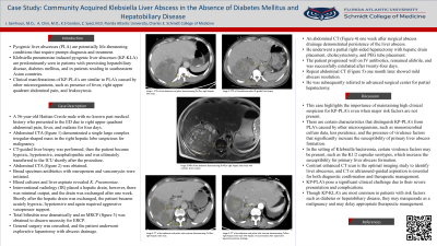
Introduction: Pyogenic liver abscesses (PLA) are potentially life-threatening conditions that require prompt diagnosis and treatment. Klebsiella pneumoniae induced pyogenic liver abscesses (KP-KLA) are predominantly seen in patients with preexisting hepatobiliary disease, diabetes mellitus, and in patients residing in southeastern Asian countries.
Case Description/Methods: A 56-year-old Haitian Creole male with no known past medical history presented to the ED due to right upper quadrant abdominal pain, fever, and malaise for four days. An abdominal computed tomography (CT) scan showed a single large complex irregular-shaped mass in the right lobe of the liver that was initially speculated to be a primary malignancy. A CT-guided liver biopsy was performed and the patient subsequently became hypoxic and hypotensive after the procedure. He was placed on mechanical ventilation and transferred to the ICU for sepsis with acute hypoxic respiratory failure. Blood and abscess cultures revealed K. Pneumoniae and broad spectrum antibiotics with meropenem and vancomycin were initiated. Interventional radiology (IR) placed a biliary catheter to allows for abscess drainage. An abdominal CT demonstrated a large liver mass with a right perihepatic hematoma, a right pleural effusion with atelectasis, and prominent small bowel loops. Surgery was consulted and the patient underwent an explorative laparotomy. He received a cholecystectomy after he was found to have a gangrenous gallbladder, a right sided thoracostomy tube and a PEG tube for nutritional feeding. The patient progressed well on IV antibiotics, remained afebrile, and was successfully extubated after twenty-four days of ventilator dependence.
Discussion: This case highlights the importance of maintaining high clinical suspicion for KP-PLA’s even when major risk factors are not present. In the setting of Klebsiella bacteremia, certain virulence factors may be present, such as the K1/2 capsular serotypes, which increase the susceptibility for primary liver abscess formation. Contrast enhanced CT scan is the optimal imaging study to identify liver abscesses, and CT or ultrasound-guided aspiration is essential for both diagnostic confirmation and therapeutic management. KP-PLA’s pose a significant clinical challenge due to their severe presentation and complications. Though KP-KLA’s are most common in patients with risk factors such as diabetes or hepatobiliary disease, they may masquerade as a malignancy and may delay appropriate therapeutic management.

Disclosures:
Jalal Samhoun, MD1, Allison Chin, MD2, Reem Ghalib, MD3, Zaineb Syed, MD3. P4869 - Case Study: Community-Acquired Klebsiella Liver Abscess in the Absence of Diabetes Mellitus and Hepatobiliary Disease, ACG 2024 Annual Scientific Meeting Abstracts. Philadelphia, PA: American College of Gastroenterology.
1Florida Atlantic University, Wellington, FL; 2Florida Atlantic University, Miami, FL; 3Florida Atlantic University, Boca Raton, FL
Introduction: Pyogenic liver abscesses (PLA) are potentially life-threatening conditions that require prompt diagnosis and treatment. Klebsiella pneumoniae induced pyogenic liver abscesses (KP-KLA) are predominantly seen in patients with preexisting hepatobiliary disease, diabetes mellitus, and in patients residing in southeastern Asian countries.
Case Description/Methods: A 56-year-old Haitian Creole male with no known past medical history presented to the ED due to right upper quadrant abdominal pain, fever, and malaise for four days. An abdominal computed tomography (CT) scan showed a single large complex irregular-shaped mass in the right lobe of the liver that was initially speculated to be a primary malignancy. A CT-guided liver biopsy was performed and the patient subsequently became hypoxic and hypotensive after the procedure. He was placed on mechanical ventilation and transferred to the ICU for sepsis with acute hypoxic respiratory failure. Blood and abscess cultures revealed K. Pneumoniae and broad spectrum antibiotics with meropenem and vancomycin were initiated. Interventional radiology (IR) placed a biliary catheter to allows for abscess drainage. An abdominal CT demonstrated a large liver mass with a right perihepatic hematoma, a right pleural effusion with atelectasis, and prominent small bowel loops. Surgery was consulted and the patient underwent an explorative laparotomy. He received a cholecystectomy after he was found to have a gangrenous gallbladder, a right sided thoracostomy tube and a PEG tube for nutritional feeding. The patient progressed well on IV antibiotics, remained afebrile, and was successfully extubated after twenty-four days of ventilator dependence.
Discussion: This case highlights the importance of maintaining high clinical suspicion for KP-PLA’s even when major risk factors are not present. In the setting of Klebsiella bacteremia, certain virulence factors may be present, such as the K1/2 capsular serotypes, which increase the susceptibility for primary liver abscess formation. Contrast enhanced CT scan is the optimal imaging study to identify liver abscesses, and CT or ultrasound-guided aspiration is essential for both diagnostic confirmation and therapeutic management. KP-PLA’s pose a significant clinical challenge due to their severe presentation and complications. Though KP-KLA’s are most common in patients with risk factors such as diabetes or hepatobiliary disease, they may masquerade as a malignancy and may delay appropriate therapeutic management.

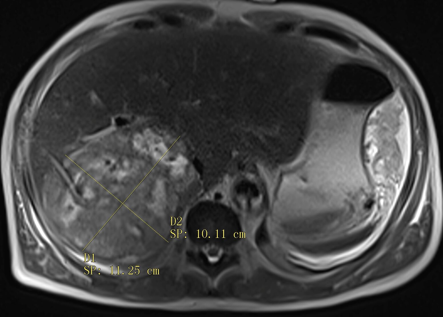
Figure: Abdominal MRI without contrast demonstrating right hepatic lobe abscess with percutaneous catheter drain in place.
Disclosures:
Jalal Samhoun indicated no relevant financial relationships.
Allison Chin indicated no relevant financial relationships.
Reem Ghalib indicated no relevant financial relationships.
Zaineb Syed indicated no relevant financial relationships.
Jalal Samhoun, MD1, Allison Chin, MD2, Reem Ghalib, MD3, Zaineb Syed, MD3. P4869 - Case Study: Community-Acquired Klebsiella Liver Abscess in the Absence of Diabetes Mellitus and Hepatobiliary Disease, ACG 2024 Annual Scientific Meeting Abstracts. Philadelphia, PA: American College of Gastroenterology.
