Tuesday Poster Session
Category: Small Intestine
P4946 - Whipple’s Disease: A Rare Endoscopic Appearance of Rare Disease
Tuesday, October 29, 2024
10:30 AM - 4:00 PM ET
Location: Exhibit Hall E

Has Audio
- CS
Claire Shin, MD
Oregon Health & Science University
Portland, OR
Presenting Author(s)
Claire Shin, MD, Brian Mau, MD, Zhengchun Lu, MD, MSc, PhD, Brian Brinkerhoff, MD, Seth Crockett, MD
Oregon Health & Science University, Portland, OR
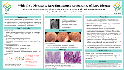
Introduction: Whipple’s disease is a rare, chronic, and systemic infectious disease that primarily impacts the gastrointestinal tract leading to abdominal pain, diarrhea, and malnutrition. We present a case of Whipple disease in the small intestine with endoscopic findings of atypical white-tipped villi with granular appearance in duodenum and terminal ileum.
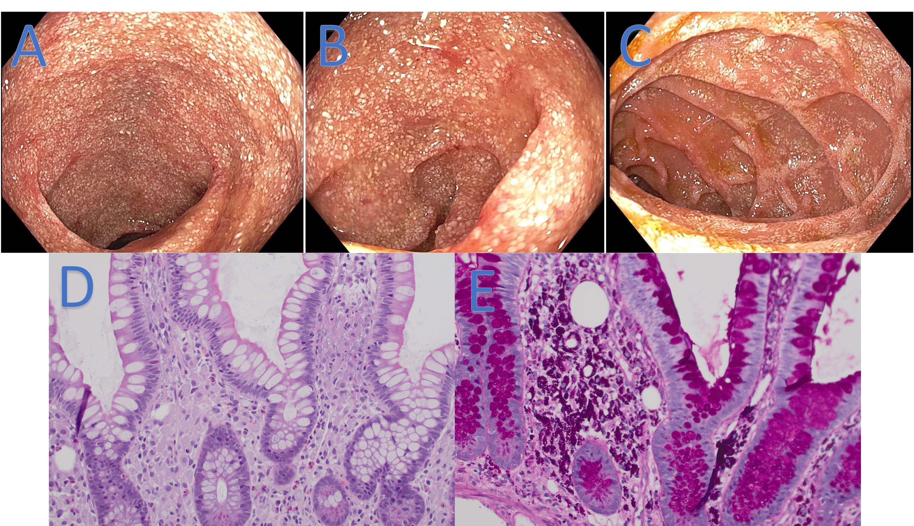
Case Description/Methods: A 46-year-old male without chronic medical conditions presented to ED with 6 months of persistent diarrhea (up to 4-6 bowel movements per day), and 40-pound weight loss. The patient had tried lactose and gluten-free diet, and loperamide and pancreatic enzyme supplementation without relief. The patient described his bowel movement as foul-smelling, greasy, and fatty. Labs revealed electrolyte derangement (K 2.4, Cl 99, CO2 29, Mg 0.5), iron deficiency anemia (Hgb 6.8, MCV 63, ferritin 10, iron 14), low albumin (2.6), and fat-soluble vitamin levels. Other testings were normal including Celiac serologies, HIV, TSH, H pylori, and stool infectious studies. CT abdomen and pelvis showed mesenteric lymphadenopathy but no active bowel inflammation. The patient underwent bidirectional endoscopy that showed atypical white-tipped villi with granular appearance in the duodenum and terminal ileum (Figure 1). The H&E stain showed numerous lamina propria foamy histiocytes in the small bowel mucosa (Figure 1). The periodic acid Schiff (PAS) stain showed positive granular cytoplasmic staining in the histiocytes, confirming the diagnosis of Whipple disease (Figure 1). T. Whipplei PCR of the duodenum and terminal ileum also confirmed the diagnosis. Lumbar puncture was negative for CNS involvement. The patient was started on 2 weeks of IV Ceftriaxone followed by Bactrim for several months, leading to the resolution of symptoms.
Discussion: We present a patient with rare Whipple’s disease with endoscopic visualization of macroscopic changes in the duodenum and terminal ileum. The disease was associated with weight loss, nutritional deficiency, iron deficiency anemia, and persistent malabsorptive diarrhea. Endoscopists should be aware of endoscopic findings of Whipple disease, which may affect distal small bowel.

Disclosures:
Claire Shin, MD, Brian Mau, MD, Zhengchun Lu, MD, MSc, PhD, Brian Brinkerhoff, MD, Seth Crockett, MD. P4946 - Whipple’s Disease: A Rare Endoscopic Appearance of Rare Disease, ACG 2024 Annual Scientific Meeting Abstracts. Philadelphia, PA: American College of Gastroenterology.
Oregon Health & Science University, Portland, OR
Introduction: Whipple’s disease is a rare, chronic, and systemic infectious disease that primarily impacts the gastrointestinal tract leading to abdominal pain, diarrhea, and malnutrition. We present a case of Whipple disease in the small intestine with endoscopic findings of atypical white-tipped villi with granular appearance in duodenum and terminal ileum.
Case Description/Methods: A 46-year-old male without chronic medical conditions presented to ED with 6 months of persistent diarrhea (up to 4-6 bowel movements per day), and 40-pound weight loss. The patient had tried lactose and gluten-free diet, and loperamide and pancreatic enzyme supplementation without relief. The patient described his bowel movement as foul-smelling, greasy, and fatty. Labs revealed electrolyte derangement (K 2.4, Cl 99, CO2 29, Mg 0.5), iron deficiency anemia (Hgb 6.8, MCV 63, ferritin 10, iron 14), low albumin (2.6), and fat-soluble vitamin levels. Other testings were normal including Celiac serologies, HIV, TSH, H pylori, and stool infectious studies. CT abdomen and pelvis showed mesenteric lymphadenopathy but no active bowel inflammation. The patient underwent bidirectional endoscopy that showed atypical white-tipped villi with granular appearance in the duodenum and terminal ileum (Figure 1). The H&E stain showed numerous lamina propria foamy histiocytes in the small bowel mucosa (Figure 1). The periodic acid Schiff (PAS) stain showed positive granular cytoplasmic staining in the histiocytes, confirming the diagnosis of Whipple disease (Figure 1). T. Whipplei PCR of the duodenum and terminal ileum also confirmed the diagnosis. Lumbar puncture was negative for CNS involvement. The patient was started on 2 weeks of IV Ceftriaxone followed by Bactrim for several months, leading to the resolution of symptoms.
Discussion: We present a patient with rare Whipple’s disease with endoscopic visualization of macroscopic changes in the duodenum and terminal ileum. The disease was associated with weight loss, nutritional deficiency, iron deficiency anemia, and persistent malabsorptive diarrhea. Endoscopists should be aware of endoscopic findings of Whipple disease, which may affect distal small bowel.

Figure: Figure 1: Endoscopic and histologic findings of Whipple’s disease
A) Duodenum: white-tipped villi with granular appearance
B&C) Terminal Ileum: white-tipped villi with granular appearance
D) Terminal ileum H&E stain: infiltration of the lamina propria by histiocytes which contain pale nucleus with abundant cytoplasm
E) Terminal ileum PAS stain: histiocytes containing PAS-positive intracytoplasmic granules
Disclosures:
Claire Shin indicated no relevant financial relationships.
Brian Mau indicated no relevant financial relationships.
Zhengchun Lu indicated no relevant financial relationships.
Brian Brinkerhoff indicated no relevant financial relationships.
Seth Crockett indicated no relevant financial relationships.
Claire Shin, MD, Brian Mau, MD, Zhengchun Lu, MD, MSc, PhD, Brian Brinkerhoff, MD, Seth Crockett, MD. P4946 - Whipple’s Disease: A Rare Endoscopic Appearance of Rare Disease, ACG 2024 Annual Scientific Meeting Abstracts. Philadelphia, PA: American College of Gastroenterology.
