Tuesday Poster Session
Category: Small Intestine
P4948 - Invasive Adenocarcinoma of the Terminal Ileum Presenting as a Small Bowel Obstruction: A Rare and Challenging Entity
Tuesday, October 29, 2024
10:30 AM - 4:00 PM ET
Location: Exhibit Hall E

Has Audio
- YA
Yasir Ali, MD
Brookdale University Hospital Medical Center
Brooklyn, NY
Presenting Author(s)
Yasir Ali, MD1, Derrick Cheung, MD2
1Brookdale University Hospital Medical Center, Brooklyn, NY; 2One Brooklyn Health-Brookdale University Hospital Medical Center, Brooklyn, NY
Introduction: Invasive adenocarcinoma of the terminal ileum (TI) is an extremely rare tumor with an annual incidence reported as less than one case per million. Due to very low prevalence, a high index of suspicion is crucial to make an early diagnosis. We report a case of invasive adenocarcinoma of TI which was diagnosed and treated prior to distant metastasis, highlighting the importance of an earlier diagnosis and standard surgical treatment for improved patient outcomes.
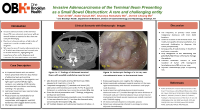
Case Description/Methods: A 63-year-old female with no medical history presented with a few days history of abdominal pain and vomiting. Reported pain mainly in the epigastric and right upper quadrant (RUQ) region, constant, non radiating, 4/10 intensity, associated with non bilious non bloody vomiting x 4-5 episodes. Last bowel movement was reported a day prior to admission. Physical exam was remarkable for an underweight patient with pale conjunctivae, mild epigastric and RUQ tenderness with sluggish bowel sounds. Vital signs were stable. Labs showed normocytic anemia, CEA level was mildly elevated to 6.3 ng/ml (n = 0 to 4.7). Computed tomography (CT) revealed small bowel (SB) obstruction with transition point at the TI. The TI appears thickened, an underlying mass cannot be excluded (Fig. 1a). Given clinical presentation and CT findings, malignancy workup was warranted. Colonoscopy with ileoscopy revealed a near circumferential, 3-4 cm mass found in the TI, 5 cm from the Ileocecal valve, concerning for SB neoplasm (Fig. 1b). S/P multiple biopsies and submucosal injection of tattoo x 2. Endoscopic biopsies were negative for malignancy. Patient eventually underwent surgical resection of the mass, right hemicolectomy with ileocolic anastomosis and lymph node dissection. Surgical specimen pathology demonstrated invasive adenocarcinoma of TI, invades through the entire thickness of the bowel wall, peri-intestinal adipose tissue and perineural involvement (no blood vessels invasion was noted) consistent with T3N1M0 staging (CT chest and head shows no metastatic process). Patient was subsequently referred to oncology to initiate adjuvant chemotherapy.
Discussion: Given its location in the distal SB and nonspecific clinical presentation, it is extremely challenging to diagnose this tumor preoperatively. Consequently, it leads to delay in treatment and a poor prognosis. Standard treatment consists of wide resection of tumor with locoregional lymphadenectomy, as in our case. Adjuvant chemotherapy plays a supporting role.

Disclosures:
Yasir Ali, MD1, Derrick Cheung, MD2. P4948 - Invasive Adenocarcinoma of the Terminal Ileum Presenting as a Small Bowel Obstruction: A Rare and Challenging Entity, ACG 2024 Annual Scientific Meeting Abstracts. Philadelphia, PA: American College of Gastroenterology.
1Brookdale University Hospital Medical Center, Brooklyn, NY; 2One Brooklyn Health-Brookdale University Hospital Medical Center, Brooklyn, NY
Introduction: Invasive adenocarcinoma of the terminal ileum (TI) is an extremely rare tumor with an annual incidence reported as less than one case per million. Due to very low prevalence, a high index of suspicion is crucial to make an early diagnosis. We report a case of invasive adenocarcinoma of TI which was diagnosed and treated prior to distant metastasis, highlighting the importance of an earlier diagnosis and standard surgical treatment for improved patient outcomes.
Case Description/Methods: A 63-year-old female with no medical history presented with a few days history of abdominal pain and vomiting. Reported pain mainly in the epigastric and right upper quadrant (RUQ) region, constant, non radiating, 4/10 intensity, associated with non bilious non bloody vomiting x 4-5 episodes. Last bowel movement was reported a day prior to admission. Physical exam was remarkable for an underweight patient with pale conjunctivae, mild epigastric and RUQ tenderness with sluggish bowel sounds. Vital signs were stable. Labs showed normocytic anemia, CEA level was mildly elevated to 6.3 ng/ml (n = 0 to 4.7). Computed tomography (CT) revealed small bowel (SB) obstruction with transition point at the TI. The TI appears thickened, an underlying mass cannot be excluded (Fig. 1a). Given clinical presentation and CT findings, malignancy workup was warranted. Colonoscopy with ileoscopy revealed a near circumferential, 3-4 cm mass found in the TI, 5 cm from the Ileocecal valve, concerning for SB neoplasm (Fig. 1b). S/P multiple biopsies and submucosal injection of tattoo x 2. Endoscopic biopsies were negative for malignancy. Patient eventually underwent surgical resection of the mass, right hemicolectomy with ileocolic anastomosis and lymph node dissection. Surgical specimen pathology demonstrated invasive adenocarcinoma of TI, invades through the entire thickness of the bowel wall, peri-intestinal adipose tissue and perineural involvement (no blood vessels invasion was noted) consistent with T3N1M0 staging (CT chest and head shows no metastatic process). Patient was subsequently referred to oncology to initiate adjuvant chemotherapy.
Discussion: Given its location in the distal SB and nonspecific clinical presentation, it is extremely challenging to diagnose this tumor preoperatively. Consequently, it leads to delay in treatment and a poor prognosis. Standard treatment consists of wide resection of tumor with locoregional lymphadenectomy, as in our case. Adjuvant chemotherapy plays a supporting role.

Figure: Fig. 1a: CT findings of thickened terminal ileum with possible underlying mass (arrow)
Fig. 1b: Endoscopic findings of near circumferential, 3-4 cm mass found in the terminal ileum
Fig. 1b: Endoscopic findings of near circumferential, 3-4 cm mass found in the terminal ileum
Disclosures:
Yasir Ali indicated no relevant financial relationships.
Derrick Cheung indicated no relevant financial relationships.
Yasir Ali, MD1, Derrick Cheung, MD2. P4948 - Invasive Adenocarcinoma of the Terminal Ileum Presenting as a Small Bowel Obstruction: A Rare and Challenging Entity, ACG 2024 Annual Scientific Meeting Abstracts. Philadelphia, PA: American College of Gastroenterology.
