Tuesday Poster Session
Category: Small Intestine
P4961 - Isolated Jejunal Varices: A Hidden Source of Gastrointestinal Bleeding in Patient With Extrahepatic Portal Hypertension and Altered Anatomy
Tuesday, October 29, 2024
10:30 AM - 4:00 PM ET
Location: Exhibit Hall E

Has Audio

Rama Tarakji, MD
Mount Sinai Medical Center
Miami Beach, FL
Presenting Author(s)
Award: Presidential Poster Award
Rama Tarakji, MD1, Rahil H Shah, MD2, Mohamed Karam Douedari, MD1, Mahmoud Mahfouz, MD2
1Mount Sinai Medical Center, Miami Beach, FL; 2University of Miami Miller School of Medicine, Miami, FL
Introduction: Ectopic varices in the small intestine are uncommon findings typically caused by portal hypertension secondary to underlying hepatic cirrhosis. Isolated jejunal variceal bleeding has been rarely described and represents a diagnostic challenge. We present a case of recurrent gastrointestinal bleeding due to isolated jejunal varices.
Case Description/Methods: A 21-year-old woman with a history of a choledochal cyst status post excision with roux-en-Y choledochojejunostomy complicated by chronic portal vein thrombosis and cavernous transformation of the portal vein resulting in portal hypertension and splenomegaly was being followed for chronic anemia with recurrent episodes of obscure gastrointestinal bleeding. She presented with melena and a hemoglobin of 3.2 g/dl. An extensive workup including Computed tomography (CT) angiography, nuclear medicine bleeding scan, push enteroscopy, and colonoscopy was unremarkable. She underwent a single balloon enteroscopy with fluoroscopic guidance and was found to have a bleeding arteriovenous malformation lesion which was successfully treated with Argon plasma coagulation. However, the patient continued to have intermittent episodes of melena for 2 months. Repeat endoscopy showed diffuse erythematous and friable mucosa suspicious of recent bleeding (Figure 1,2). Biopsies obtained from the area resulted in active bleeding that was successfully controlled with epinephrine and hemostatic spray. CT confirmed the diagnosis of isolated jejunal varices (Figure 3,4).
Discussion: Isolated jejunal varices represent a rare source of recurrent gastrointestinal bleeding in patients with portal hypertension. The diagnosis is often delayed, requiring the patient to undergo multiple procedures. In our case, the diagnosis was also difficult due to the complex anatomy from the previous Roux-en-Y choledochojejunostomy. Kohli et al. described a case of a man with a history of liver transplant with Roux-en-Y choledochojejunostomy and persistent portal hypertension who presented with anemia and was found to have jejunal varices at the choledochojejunal anastomosis on abdominal CT. Our case adds to the limited reports of isolated jejunal varices in patients with extrahepatic portal hypertension and altered anatomy. It also highlights the importance of considering isolated jejunal varices in the differential diagnosis of gastrointestinal bleeding in these patients, as early recognition can prevent multiple procedures and reduce the risk of life-threatening and recurrent bleeding.

Disclosures:
Rama Tarakji, MD1, Rahil H Shah, MD2, Mohamed Karam Douedari, MD1, Mahmoud Mahfouz, MD2. P4961 - Isolated Jejunal Varices: A Hidden Source of Gastrointestinal Bleeding in Patient With Extrahepatic Portal Hypertension and Altered Anatomy, ACG 2024 Annual Scientific Meeting Abstracts. Philadelphia, PA: American College of Gastroenterology.
Rama Tarakji, MD1, Rahil H Shah, MD2, Mohamed Karam Douedari, MD1, Mahmoud Mahfouz, MD2
1Mount Sinai Medical Center, Miami Beach, FL; 2University of Miami Miller School of Medicine, Miami, FL
Introduction: Ectopic varices in the small intestine are uncommon findings typically caused by portal hypertension secondary to underlying hepatic cirrhosis. Isolated jejunal variceal bleeding has been rarely described and represents a diagnostic challenge. We present a case of recurrent gastrointestinal bleeding due to isolated jejunal varices.
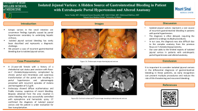
Case Description/Methods: A 21-year-old woman with a history of a choledochal cyst status post excision with roux-en-Y choledochojejunostomy complicated by chronic portal vein thrombosis and cavernous transformation of the portal vein resulting in portal hypertension and splenomegaly was being followed for chronic anemia with recurrent episodes of obscure gastrointestinal bleeding. She presented with melena and a hemoglobin of 3.2 g/dl. An extensive workup including Computed tomography (CT) angiography, nuclear medicine bleeding scan, push enteroscopy, and colonoscopy was unremarkable. She underwent a single balloon enteroscopy with fluoroscopic guidance and was found to have a bleeding arteriovenous malformation lesion which was successfully treated with Argon plasma coagulation. However, the patient continued to have intermittent episodes of melena for 2 months. Repeat endoscopy showed diffuse erythematous and friable mucosa suspicious of recent bleeding (Figure 1,2). Biopsies obtained from the area resulted in active bleeding that was successfully controlled with epinephrine and hemostatic spray. CT confirmed the diagnosis of isolated jejunal varices (Figure 3,4).
Discussion: Isolated jejunal varices represent a rare source of recurrent gastrointestinal bleeding in patients with portal hypertension. The diagnosis is often delayed, requiring the patient to undergo multiple procedures. In our case, the diagnosis was also difficult due to the complex anatomy from the previous Roux-en-Y choledochojejunostomy. Kohli et al. described a case of a man with a history of liver transplant with Roux-en-Y choledochojejunostomy and persistent portal hypertension who presented with anemia and was found to have jejunal varices at the choledochojejunal anastomosis on abdominal CT. Our case adds to the limited reports of isolated jejunal varices in patients with extrahepatic portal hypertension and altered anatomy. It also highlights the importance of considering isolated jejunal varices in the differential diagnosis of gastrointestinal bleeding in these patients, as early recognition can prevent multiple procedures and reduce the risk of life-threatening and recurrent bleeding.

Figure: Figure 1 and 2. Endoscopic image showing isolated jejunal varices
Figure 3 and 4. Contrast-enhanced CT scan image revealing isolated jejunal varices
Figure 3 and 4. Contrast-enhanced CT scan image revealing isolated jejunal varices
Disclosures:
Rama Tarakji indicated no relevant financial relationships.
Rahil H Shah indicated no relevant financial relationships.
Mohamed Karam Douedari indicated no relevant financial relationships.
Mahmoud Mahfouz indicated no relevant financial relationships.
Rama Tarakji, MD1, Rahil H Shah, MD2, Mohamed Karam Douedari, MD1, Mahmoud Mahfouz, MD2. P4961 - Isolated Jejunal Varices: A Hidden Source of Gastrointestinal Bleeding in Patient With Extrahepatic Portal Hypertension and Altered Anatomy, ACG 2024 Annual Scientific Meeting Abstracts. Philadelphia, PA: American College of Gastroenterology.

