Tuesday Poster Session
Category: Small Intestine
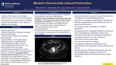
P4964 - Meckel’s Diverticulitis-Induced Perforation
Tuesday, October 29, 2024
10:30 AM - 4:00 PM ET
Location: Exhibit Hall E

Has Audio
- RP
Rahul Patel, DO
Jefferson Health
Washington Township, NJ
Presenting Author(s)
Rahul Patel, DO1, Edward Bley, DO1, Joshua Soliman, DO1, Charles Kistler, MD2
1Jefferson Health, Washington Township, NJ; 2Sidney Kimmel Medical College at Thomas Jefferson University, Philadelphia, PA
Introduction: Meckel’s Diverticulum is a vestigial remnant seen in 0.3-3% of the population. We present an unusual case of an elderly female with perforation in the setting of Meckel’s diverticulitis.
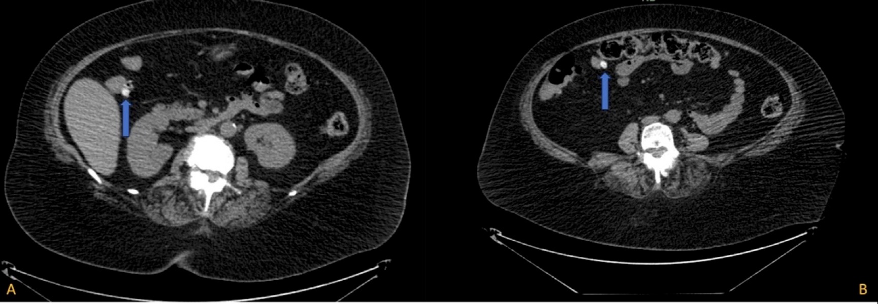
Case Description/Methods: A 66-year-old female with a relevant history of medically managed recurrent diverticulitis presented to the hospital with right lower quadrant abdominal pain. Surgical history was significant for remote laparoscopic cholecystectomy and appendectomy. She arrived hemodynamically stable. Her lab work showed a leukocytosis of 16.2 B/L with normal hepatic panel and lipase. A computerized tomography (CT) of the abdomen and pelvis without contrast (figure 1) was done showing an elongated blind-ending tubular structure arising from distal small bowel in the right abdomen with a calcification near its terminus suggestive of diverticulitis involving Meckel’s diverticulum with a calculus near the terminus of the diverticulum. She underwent exploratory laparoscopy for intended diverticulectomy and small bowel resection. Intraoperatively, perforation of the small bowel with phlegmon in the right lower quadrant with purulent peritonitis was observed. The segment before and after perforation was resected and adhesions were lysed. The resected margins were biopsied and showed acute inflammation without necrosis/malignancy. Post operatively, she was managed with a nasogastric tube, intravenous ciprofloxacin and metronidazole for a 5-day duration.
Discussion: Meckel's diverticulitis is rare to the point that there are no statistics on its incidence/prevalence. Less than 2% of patients with Meckel’s diverticulum develop complications and the findings are rarer in women and the elderly The most common complication in adults is obstruction. Our patient had an undetected Meckel’s diverticulum with complications of enterolith formation and recurrent diverticulitis with subsequent perforation. Enteroliths only form in 3-10% of Meckel’s diverticulum due to bowel stasis. They are rarely seen on imaging. Our patient’s perforation was likely from the diverticulitis but the observation of the enterolith was unique. Despite the rarity of Meckel's diverticulitis, this case also highlighted the treatment of choice for symptomatic Meckel’s diverticulum being surgical resection.

Disclosures:
Rahul Patel, DO1, Edward Bley, DO1, Joshua Soliman, DO1, Charles Kistler, MD2. P4964 - Meckel’s Diverticulitis-Induced Perforation, ACG 2024 Annual Scientific Meeting Abstracts. Philadelphia, PA: American College of Gastroenterology.
1Jefferson Health, Washington Township, NJ; 2Sidney Kimmel Medical College at Thomas Jefferson University, Philadelphia, PA
Introduction: Meckel’s Diverticulum is a vestigial remnant seen in 0.3-3% of the population. We present an unusual case of an elderly female with perforation in the setting of Meckel’s diverticulitis.
Case Description/Methods: A 66-year-old female with a relevant history of medically managed recurrent diverticulitis presented to the hospital with right lower quadrant abdominal pain. Surgical history was significant for remote laparoscopic cholecystectomy and appendectomy. She arrived hemodynamically stable. Her lab work showed a leukocytosis of 16.2 B/L with normal hepatic panel and lipase. A computerized tomography (CT) of the abdomen and pelvis without contrast (figure 1) was done showing an elongated blind-ending tubular structure arising from distal small bowel in the right abdomen with a calcification near its terminus suggestive of diverticulitis involving Meckel’s diverticulum with a calculus near the terminus of the diverticulum. She underwent exploratory laparoscopy for intended diverticulectomy and small bowel resection. Intraoperatively, perforation of the small bowel with phlegmon in the right lower quadrant with purulent peritonitis was observed. The segment before and after perforation was resected and adhesions were lysed. The resected margins were biopsied and showed acute inflammation without necrosis/malignancy. Post operatively, she was managed with a nasogastric tube, intravenous ciprofloxacin and metronidazole for a 5-day duration.
Discussion: Meckel's diverticulitis is rare to the point that there are no statistics on its incidence/prevalence. Less than 2% of patients with Meckel’s diverticulum develop complications and the findings are rarer in women and the elderly The most common complication in adults is obstruction. Our patient had an undetected Meckel’s diverticulum with complications of enterolith formation and recurrent diverticulitis with subsequent perforation. Enteroliths only form in 3-10% of Meckel’s diverticulum due to bowel stasis. They are rarely seen on imaging. Our patient’s perforation was likely from the diverticulitis but the observation of the enterolith was unique. Despite the rarity of Meckel's diverticulitis, this case also highlighted the treatment of choice for symptomatic Meckel’s diverticulum being surgical resection.

Figure: Figure 1: A & B both demonstrating calcification near terminus of the blind-ending tubular structure (Meckel’s enterolith within diverticulum)
Disclosures:
Rahul Patel indicated no relevant financial relationships.
Edward Bley indicated no relevant financial relationships.
Joshua Soliman indicated no relevant financial relationships.
Charles Kistler indicated no relevant financial relationships.
Rahul Patel, DO1, Edward Bley, DO1, Joshua Soliman, DO1, Charles Kistler, MD2. P4964 - Meckel’s Diverticulitis-Induced Perforation, ACG 2024 Annual Scientific Meeting Abstracts. Philadelphia, PA: American College of Gastroenterology.
