Tuesday Poster Session
Category: Small Intestine
P4973 - Kaposi Sarcoma With Diffuse Small Bowel Involvement in a Patient Living With AIDS: A Case Report
Tuesday, October 29, 2024
10:30 AM - 4:00 PM ET
Location: Exhibit Hall E

Has Audio

Wesley Chow, MD
Scripps Mercy Hospital
San Diego, CA
Presenting Author(s)
Wesley Chow, MD1, Joy-Marie Hermes, MD2, Nabil El Hage Chehade, MD3, Faizi Hai, MD1, Andrew Cummins, MD1
1Scripps Mercy Hospital, San Diego, CA; 2Scripps Clinic & Scripps Research Institute, San Diego, CA; 3Scripps Clinic, San Diego, CA
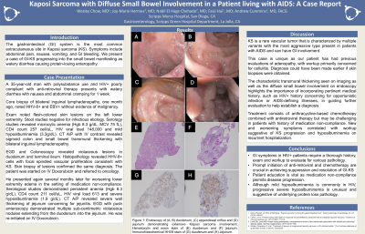
Introduction: The gastrointestinal (GI) system is the most commonly involved extracutaneous site in Kaposi sarcoma (KS). Typical manifestations include abdominal pain, nausea, vomiting, and GI bleeding. We present a case of diffuse small bowel KS manifesting as severe acute watery diarrhea causing protein-losing enteropathy.
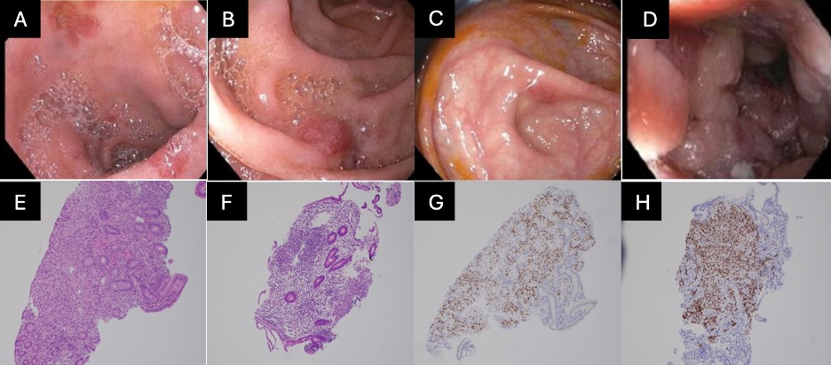
Case Description/Methods: A 35-year-old man with polysubstance use disorder and HIV poorly compliant with anti-retroviral therapy was hospitalized with acute watery diarrhea for 1 week duration and associated with nausea and abdominal cramping. Chart review revealed he had bilateral inguinal lymphadenopathy for which a prior core biopsy demonstrated cells that are positive for HHV-8 and EBV without evidence of malignancy. Physical exam showed flesh-colored skin lesions on the left lower extremity. Stool studies were negative for infection. Labs revealed hypoalbuminemia (1.9 g/dL), microcytic anemia (Hgb 8.3 g/dL, MCV 75 fL), CD4 count of 211 cell/uL, and viral load of 148,000. CT abdomen/pelvis with IV contrast revealed sigmoid colon and small bowel transmural thickening as well as chronic bilateral inguinal lymphadenopathy. EGD with push enteroscopy demonstrated multiple sub-centimetric violaceous nodules extending from the duodenum into the jejunum. Colonoscopy was pertinent for similar nodules in the terminal ileum but was otherwise normal. Histopathology revealed HHV-8 positive cells with focal spindled vascular proliferation consistent with KS. Skin biopsy of lesions confirmed the same diagnosis. As a result, the patient was initiated on IV Doxorubicin with planned oncology follow-up.
Discussion: KS is a rare vascular tumor that is characterized by multiple variants with the most aggressive type present in patients with AIDS and has GI involvement. This case is unique as our patient has had previous evaluations of adenopathy which could have made the diagnosis earlier if skin or endoscopic biopsies were obtained. The characteristic transmural thickening seen on imaging as well as the diffuse small bowel involvement on endoscopy highlights the importance of incorporating pertinent medical history in guiding further evaluation to help establish a diagnosis. New and worsening symptoms correlated with workup suggestive of KS progression on recurrent hospitalization. Treatment consists of anthracycline-based chemotherapy but may be challenging in patients with history of medication non-compliance.

Disclosures:
Wesley Chow, MD1, Joy-Marie Hermes, MD2, Nabil El Hage Chehade, MD3, Faizi Hai, MD1, Andrew Cummins, MD1. P4973 - Kaposi Sarcoma With Diffuse Small Bowel Involvement in a Patient Living With AIDS: A Case Report, ACG 2024 Annual Scientific Meeting Abstracts. Philadelphia, PA: American College of Gastroenterology.
1Scripps Mercy Hospital, San Diego, CA; 2Scripps Clinic & Scripps Research Institute, San Diego, CA; 3Scripps Clinic, San Diego, CA
Introduction: The gastrointestinal (GI) system is the most commonly involved extracutaneous site in Kaposi sarcoma (KS). Typical manifestations include abdominal pain, nausea, vomiting, and GI bleeding. We present a case of diffuse small bowel KS manifesting as severe acute watery diarrhea causing protein-losing enteropathy.
Case Description/Methods: A 35-year-old man with polysubstance use disorder and HIV poorly compliant with anti-retroviral therapy was hospitalized with acute watery diarrhea for 1 week duration and associated with nausea and abdominal cramping. Chart review revealed he had bilateral inguinal lymphadenopathy for which a prior core biopsy demonstrated cells that are positive for HHV-8 and EBV without evidence of malignancy. Physical exam showed flesh-colored skin lesions on the left lower extremity. Stool studies were negative for infection. Labs revealed hypoalbuminemia (1.9 g/dL), microcytic anemia (Hgb 8.3 g/dL, MCV 75 fL), CD4 count of 211 cell/uL, and viral load of 148,000. CT abdomen/pelvis with IV contrast revealed sigmoid colon and small bowel transmural thickening as well as chronic bilateral inguinal lymphadenopathy. EGD with push enteroscopy demonstrated multiple sub-centimetric violaceous nodules extending from the duodenum into the jejunum. Colonoscopy was pertinent for similar nodules in the terminal ileum but was otherwise normal. Histopathology revealed HHV-8 positive cells with focal spindled vascular proliferation consistent with KS. Skin biopsy of lesions confirmed the same diagnosis. As a result, the patient was initiated on IV Doxorubicin with planned oncology follow-up.
Discussion: KS is a rare vascular tumor that is characterized by multiple variants with the most aggressive type present in patients with AIDS and has GI involvement. This case is unique as our patient has had previous evaluations of adenopathy which could have made the diagnosis earlier if skin or endoscopic biopsies were obtained. The characteristic transmural thickening seen on imaging as well as the diffuse small bowel involvement on endoscopy highlights the importance of incorporating pertinent medical history in guiding further evaluation to help establish a diagnosis. New and worsening symptoms correlated with workup suggestive of KS progression on recurrent hospitalization. Treatment consists of anthracycline-based chemotherapy but may be challenging in patients with history of medication non-compliance.

Figure: Figure 1: Endoscopic appearance of (A, B) duodenum, (C) appendiceal orifice and (D) jejunum demonstrating extensive Kaposi sarcoma involvement. Hematoxylin and eosin stain of (E) duodenum and (F) jejunum. Immunohistochemical HHV8 stain of (G) duodenum and (H) Jejunum.
Disclosures:
Wesley Chow indicated no relevant financial relationships.
Joy-Marie Hermes indicated no relevant financial relationships.
Nabil El Hage Chehade indicated no relevant financial relationships.
Faizi Hai indicated no relevant financial relationships.
Andrew Cummins indicated no relevant financial relationships.
Wesley Chow, MD1, Joy-Marie Hermes, MD2, Nabil El Hage Chehade, MD3, Faizi Hai, MD1, Andrew Cummins, MD1. P4973 - Kaposi Sarcoma With Diffuse Small Bowel Involvement in a Patient Living With AIDS: A Case Report, ACG 2024 Annual Scientific Meeting Abstracts. Philadelphia, PA: American College of Gastroenterology.
