Tuesday Poster Session
Category: Small Intestine
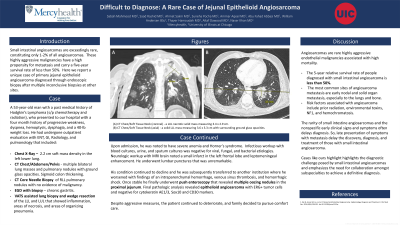
P4981 - Difficult to Diagnose: A Rare Case of Jejunal Epithelioid Angiosarcoma
Tuesday, October 29, 2024
10:30 AM - 4:00 PM ET
Location: Exhibit Hall E

Has Audio
- SM
Sabah Mahmood, MD
Mercy Health
Rockford, IL
Presenting Author(s)
Sabah Mahmood, MD1, Saad Rashid, MD1, Ahmet Sakiri, MD1, Suneha Pocha, MD1, Ammar Aqeel, MD2, Abu Fahad Abbasi, MD1, William Andersen, BSc3, Thayer Hamoudah, MD1, Altaf Dawood, MD, MBBS1, Naser Khan, MD1
1Mercy Health, Rockford, IL; 2Mercy Health, Loves Park, IL; 3University of Illinois College of Medicine, Loves Park, IL
Introduction: Small intestinal angiosarcomas are exceedingly rare, constituting only 1-2% of all angiosarcomas. These highly aggressive malignancies have a high propensity for metastasis and carry a five-year survival rate of less than 50%. Here we report a unique case of primary jejunal epithelioid angiosarcoma diagnosed through endoscopic biopsy after multiple inconclusive biopsies at other sites.
Case Description/Methods: A 53-year-old male with a past medical history of Hodgkin’s Lymphoma, previously treated with chemotherapy and radiation, who presented with a four-month history of progressive weakness, dyspnea, hemoptysis, dysphagia, and a significant weight loss of forty pounds. He had undergone outpatient evaluation by multiple specialties including GI, Radiology, ENT, and Pulmonology. Initial work-up included CT Chest/Abdomen/Pelvis, which revealed multiple lung masses, pulmonary nodules, and sigmoid colon thickening. A lung needle biopsy was unrevealing for malignancy. An EGD showed findings consistent with gastritis. Upon admission, repeat CT Chest showed numerous enlarging pulmonary nodules with ground glass opacities. Cardiothoracic Surgery was consulted, and Video-Assisted Thoracoscopic Surgery (VATS)lung biopsy revealed inflammatory changes but no definitive malignancy. Given the persistent clinical suspicion and lack of a clear diagnosis, a Push Enteroscopy was performed, revealing multiple oozing nodules in the proximal jejunum. Biopsies taken from these nodules revealed epithelioid angiosarcoma with tumor cells positive for ERG and negative for cytokeratin AE1/3, Sox10 and CD30 markers. Despite aggressive measures, the patient’s condition continued to deteriorate, and he was transitioned to comfort care following the biopsy results.
Discussion: Angiosarcomas are rare endothelial malignancies associated with high mortality. They demonstrate early nodal and solid organ metastasis, especially to the lungs and bone. Significant risk factors associated with angiosarcoma include prior radiation exposure and environmental toxins. The rarity of small intestine angiosarcomas and the nonspecific early clinical signs and symptoms often delays diagnosis. This case highlights the diagnostic challenge posed by small intestinal angiosarcomas and emphasizes the need for collaboration amongst subspecialties to achieve a definitive diagnosis.

Disclosures:
Sabah Mahmood, MD1, Saad Rashid, MD1, Ahmet Sakiri, MD1, Suneha Pocha, MD1, Ammar Aqeel, MD2, Abu Fahad Abbasi, MD1, William Andersen, BSc3, Thayer Hamoudah, MD1, Altaf Dawood, MD, MBBS1, Naser Khan, MD1. P4981 - Difficult to Diagnose: A Rare Case of Jejunal Epithelioid Angiosarcoma, ACG 2024 Annual Scientific Meeting Abstracts. Philadelphia, PA: American College of Gastroenterology.
1Mercy Health, Rockford, IL; 2Mercy Health, Loves Park, IL; 3University of Illinois College of Medicine, Loves Park, IL
Introduction: Small intestinal angiosarcomas are exceedingly rare, constituting only 1-2% of all angiosarcomas. These highly aggressive malignancies have a high propensity for metastasis and carry a five-year survival rate of less than 50%. Here we report a unique case of primary jejunal epithelioid angiosarcoma diagnosed through endoscopic biopsy after multiple inconclusive biopsies at other sites.
Case Description/Methods: A 53-year-old male with a past medical history of Hodgkin’s Lymphoma, previously treated with chemotherapy and radiation, who presented with a four-month history of progressive weakness, dyspnea, hemoptysis, dysphagia, and a significant weight loss of forty pounds. He had undergone outpatient evaluation by multiple specialties including GI, Radiology, ENT, and Pulmonology. Initial work-up included CT Chest/Abdomen/Pelvis, which revealed multiple lung masses, pulmonary nodules, and sigmoid colon thickening. A lung needle biopsy was unrevealing for malignancy. An EGD showed findings consistent with gastritis. Upon admission, repeat CT Chest showed numerous enlarging pulmonary nodules with ground glass opacities. Cardiothoracic Surgery was consulted, and Video-Assisted Thoracoscopic Surgery (VATS)lung biopsy revealed inflammatory changes but no definitive malignancy. Given the persistent clinical suspicion and lack of a clear diagnosis, a Push Enteroscopy was performed, revealing multiple oozing nodules in the proximal jejunum. Biopsies taken from these nodules revealed epithelioid angiosarcoma with tumor cells positive for ERG and negative for cytokeratin AE1/3, Sox10 and CD30 markers. Despite aggressive measures, the patient’s condition continued to deteriorate, and he was transitioned to comfort care following the biopsy results.
Discussion: Angiosarcomas are rare endothelial malignancies associated with high mortality. They demonstrate early nodal and solid organ metastasis, especially to the lungs and bone. Significant risk factors associated with angiosarcoma include prior radiation exposure and environmental toxins. The rarity of small intestine angiosarcomas and the nonspecific early clinical signs and symptoms often delays diagnosis. This case highlights the diagnostic challenge posed by small intestinal angiosarcomas and emphasizes the need for collaboration amongst subspecialties to achieve a definitive diagnosis.

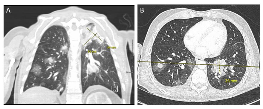
Figure: CT Chest/Soft Tissue Neck – A (Coronal) – a LUL necrotic solid mass measuring 3.4 x 2.9 cm. B (Axial) a solid LLL mass measuring 3.6 x 3.3 cm with surrounding ground glass opacities.
Disclosures:
Sabah Mahmood indicated no relevant financial relationships.
Saad Rashid indicated no relevant financial relationships.
Ahmet Sakiri indicated no relevant financial relationships.
Suneha Pocha indicated no relevant financial relationships.
Ammar Aqeel indicated no relevant financial relationships.
Abu Fahad Abbasi indicated no relevant financial relationships.
William Andersen indicated no relevant financial relationships.
Thayer Hamoudah indicated no relevant financial relationships.
Altaf Dawood indicated no relevant financial relationships.
Naser Khan indicated no relevant financial relationships.
Sabah Mahmood, MD1, Saad Rashid, MD1, Ahmet Sakiri, MD1, Suneha Pocha, MD1, Ammar Aqeel, MD2, Abu Fahad Abbasi, MD1, William Andersen, BSc3, Thayer Hamoudah, MD1, Altaf Dawood, MD, MBBS1, Naser Khan, MD1. P4981 - Difficult to Diagnose: A Rare Case of Jejunal Epithelioid Angiosarcoma, ACG 2024 Annual Scientific Meeting Abstracts. Philadelphia, PA: American College of Gastroenterology.
