Tuesday Poster Session
Category: Small Intestine
P4982 - The Path to Small Bowel Intussusception
Tuesday, October 29, 2024
10:30 AM - 4:00 PM ET
Location: Exhibit Hall E

Has Audio
- SP
Sanjay Prasad, MD
Baylor Scott & White Medical Center
Presenting Author(s)
Sanjay Prasad, MD1, Neel Shah, MD2, Manuel Garza, MD, MS2, Yizhong Wu, MD1, Douglas Larsen, MD3, Chiranjeevi Gadiparthi, MD4
1Baylor Scott & White Medical Center, Georgetown, TX; 2Baylor Scott & White Medical Center, Round Rock, TX; 3Baylor Scott & White Medical Center, Temple, TX; 4Baylor Scott and White, Round Rock, TX
Introduction: Small bowel intussusception often presents with a pathologic lead point causing intermittent abdominal pain and nausea, though it is rare in adults. We present a patient with intractable nausea and generalized abdominal pain, found to have computed tomography (CT) scan findings of jejunal intussusception later determined to be due to a large jejunal polyp.
Case Description/Methods: An 18-year-old female with no significant past medical history presented with 10 days of nausea, bilious emesis, and generalized abdominal pain, without adequate response to proton-pump inhibitors or anti-emetics at home. Her vital signs were stable, and her physical exam was significant for generalized abdominal tenderness. Serologic evaluation was unremarkable, and a computed tomography scan revealed incidental jejuno-jejunal intussusception without obstruction. The patient was treated supportively and underwent an esophagogastroduodenoscopy with push enteroscopy for further evaluation due to persistent symptoms. A 3.5 cm Paris 1S polyp was visualized in the proximal jejunum and resected with underwater endoscopic mucosal resection technique. The patient subsequently exhibited marked clinical improvement, with pathology of the resected lesion showing benign polypoid heterotopic gastric tissue with hyperplastic changes (negative for dysplasia).
Discussion: Intussusception in adults is often difficult to diagnosis given non-specific presenting symptoms, though it should be considered when the more frequent etiologies of abdominal pain have been ruled out. The overall incidence of intussusception in adults is 2-3 cases per million of the general population per year, with adult cases caused by a pathologic lead point (neoplasm most often) in 80-90% of symptomatic patients (1). A CT scan is the gold standard diagnostic tool, which has led to early diagnosis with successful nonoperative management. Our management strategy involved the use of push enteroscopy with endoscopic mucosal resection of the lead point and spared the patient surgical intervention. This case highlights the consideration of intussusception in the differential diagnoses of a young patient with non-specific abdominal pain, along with endoscopic techniques to treat intraluminal lead points of adult intussusception.
References:
(1) Panzera F, Di Venere B, Rizzi M, Biscaglia A, Praticò CA, Nasti G, Mardighian A, Nunes TF, Inchingolo R. Bowel intussusception in adult: Prevalence, diagnostic tools and therapy. World J Methodol. 2021 May 20;11(3):81-87.

Disclosures:
Sanjay Prasad, MD1, Neel Shah, MD2, Manuel Garza, MD, MS2, Yizhong Wu, MD1, Douglas Larsen, MD3, Chiranjeevi Gadiparthi, MD4. P4982 - The Path to Small Bowel Intussusception, ACG 2024 Annual Scientific Meeting Abstracts. Philadelphia, PA: American College of Gastroenterology.
1Baylor Scott & White Medical Center, Georgetown, TX; 2Baylor Scott & White Medical Center, Round Rock, TX; 3Baylor Scott & White Medical Center, Temple, TX; 4Baylor Scott and White, Round Rock, TX
Introduction: Small bowel intussusception often presents with a pathologic lead point causing intermittent abdominal pain and nausea, though it is rare in adults. We present a patient with intractable nausea and generalized abdominal pain, found to have computed tomography (CT) scan findings of jejunal intussusception later determined to be due to a large jejunal polyp.
Case Description/Methods: An 18-year-old female with no significant past medical history presented with 10 days of nausea, bilious emesis, and generalized abdominal pain, without adequate response to proton-pump inhibitors or anti-emetics at home. Her vital signs were stable, and her physical exam was significant for generalized abdominal tenderness. Serologic evaluation was unremarkable, and a computed tomography scan revealed incidental jejuno-jejunal intussusception without obstruction. The patient was treated supportively and underwent an esophagogastroduodenoscopy with push enteroscopy for further evaluation due to persistent symptoms. A 3.5 cm Paris 1S polyp was visualized in the proximal jejunum and resected with underwater endoscopic mucosal resection technique. The patient subsequently exhibited marked clinical improvement, with pathology of the resected lesion showing benign polypoid heterotopic gastric tissue with hyperplastic changes (negative for dysplasia).
Discussion: Intussusception in adults is often difficult to diagnosis given non-specific presenting symptoms, though it should be considered when the more frequent etiologies of abdominal pain have been ruled out. The overall incidence of intussusception in adults is 2-3 cases per million of the general population per year, with adult cases caused by a pathologic lead point (neoplasm most often) in 80-90% of symptomatic patients (1). A CT scan is the gold standard diagnostic tool, which has led to early diagnosis with successful nonoperative management. Our management strategy involved the use of push enteroscopy with endoscopic mucosal resection of the lead point and spared the patient surgical intervention. This case highlights the consideration of intussusception in the differential diagnoses of a young patient with non-specific abdominal pain, along with endoscopic techniques to treat intraluminal lead points of adult intussusception.
References:
(1) Panzera F, Di Venere B, Rizzi M, Biscaglia A, Praticò CA, Nasti G, Mardighian A, Nunes TF, Inchingolo R. Bowel intussusception in adult: Prevalence, diagnostic tools and therapy. World J Methodol. 2021 May 20;11(3):81-87.

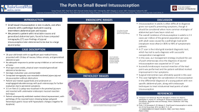
Figure: A) Polyp visualized on endoscopy
B) Underwater endoscopic mucosal resection of polyp on endoscopy
C) CT demonstrating intussusception in this case
D) 0.5x scan showing polypoid appearance of lesion
E) 10x scan demonstrating benign gastric oxyntic mucosa with mixture of chief and parietal cells; no dysplasia or malignancy visualized
Disclosures:
Sanjay Prasad indicated no relevant financial relationships.
Neel Shah indicated no relevant financial relationships.
Manuel Garza indicated no relevant financial relationships.
Yizhong Wu indicated no relevant financial relationships.
Douglas Larsen indicated no relevant financial relationships.
Chiranjeevi Gadiparthi indicated no relevant financial relationships.
Sanjay Prasad, MD1, Neel Shah, MD2, Manuel Garza, MD, MS2, Yizhong Wu, MD1, Douglas Larsen, MD3, Chiranjeevi Gadiparthi, MD4. P4982 - The Path to Small Bowel Intussusception, ACG 2024 Annual Scientific Meeting Abstracts. Philadelphia, PA: American College of Gastroenterology.
