Tuesday Poster Session
Category: Small Intestine
P5002 - Renal Cell Carcinoma: An Unusual Presentation with Melena
Tuesday, October 29, 2024
10:30 AM - 4:00 PM ET
Location: Exhibit Hall E

Has Audio

David Weinstein, MD
University of Chicago, NorthShore Internal Medicine
Evanston, IL
Presenting Author(s)
David Weinstein, MD1, Jeremy Van, DO2, Robert Toelke, MD3, Mitchelle V. Zolotarevsky, MD4, Michael Sprang, MD3
1University of Chicago, NorthShore Internal Medicine, Evanston, IL; 2University of Chicago, NorthShore University Health Systems, Chicago, IL; 3NorthShore University HealthSystem, Evanston, IL; 4University of Chicago, NorthShore Internal Medicine, Chicago, IL
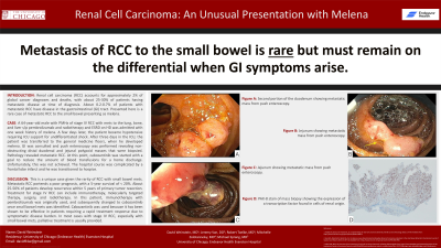
Introduction: Renal cell carcinoma (RCC) accounts for approximately 2% of global cancer diagnoses and deaths, with about 25-30% of patients having metastatic disease at time of diagnosis. The most common sites of metastases (mets) include the lung, bone, liver, and brain. About 0.2-0.7% of patients with metastatic RCC have disease in the gastrointestinal (GI) tract. Presented here is a rare case of metastatic RCC to the small bowel presenting as melena.
Case Description/Methods: A 64-year-old male with PMHx of stage IV RCC with mets to the lung, bone, and liver s/p pembrolizumab and radiotherapy and ESRD on HD was admitted with one week history of melena. On arrival, the patient was found to have community acquired pneumonia. Lab studies showed a Hgb 6.6 g/dL, WBC 15.4 x 10³/mL, Plt 210 x 103/mL, INR 1.2, Bun 38 mg/dL, and Cr 7.4 mg/dL. DRE revealed brown stool with a stable Hgb after 2 pRBC’s. A few days later, the patient became hypotensive requiring ICU support for undifferentiated shock. After three days in the ICU, the patient was transferred to the general medicine floors, when he developed melena. GI was consulted and push enteroscopy was performed revealing non-obstructing distal duodenal and jejunal polypoid masses that were biopsied. Pathology revealed metastatic RCC. At this point, cabozantinib was started with a goal to reduce the amount of blood transfusions for a home discharge. Unfortunately, this was not achieved. The hospital course was complicated by a frontal lobe infarct and he was transitioned to hospice.
Discussion: This is a unique case given the rarity of RCC with small bowel mets. Metastatic RCC portends a poor prognosis, with a 5-year survival of < 20%. Small bowel mets can present as hematochezia, melena, anemia, fatigue, weight loss, obstruction, or perforation. About 25-50% of patients develop recurrence within 5 years of primary tumor resection. Treatment for stage IV RCC can include immunotherapy, molecularly targeted therapy, surgery, and radiotherapy. In this patient, immunotherapy with pembrolizumab was originally used, and subsequently changed to cabozantinib once small bowel mets was identified. Cabozantinib was used because it has been shown to be effective in patients requiring a rapid treatment response due to symptomatic disease burden. In most cases with stage IV RCC, especially with small bowel mets, palliative treatment is usually provided.

Disclosures:
David Weinstein, MD1, Jeremy Van, DO2, Robert Toelke, MD3, Mitchelle V. Zolotarevsky, MD4, Michael Sprang, MD3. P5002 - Renal Cell Carcinoma: An Unusual Presentation with Melena, ACG 2024 Annual Scientific Meeting Abstracts. Philadelphia, PA: American College of Gastroenterology.
1University of Chicago, NorthShore Internal Medicine, Evanston, IL; 2University of Chicago, NorthShore University Health Systems, Chicago, IL; 3NorthShore University HealthSystem, Evanston, IL; 4University of Chicago, NorthShore Internal Medicine, Chicago, IL
Introduction: Renal cell carcinoma (RCC) accounts for approximately 2% of global cancer diagnoses and deaths, with about 25-30% of patients having metastatic disease at time of diagnosis. The most common sites of metastases (mets) include the lung, bone, liver, and brain. About 0.2-0.7% of patients with metastatic RCC have disease in the gastrointestinal (GI) tract. Presented here is a rare case of metastatic RCC to the small bowel presenting as melena.
Case Description/Methods: A 64-year-old male with PMHx of stage IV RCC with mets to the lung, bone, and liver s/p pembrolizumab and radiotherapy and ESRD on HD was admitted with one week history of melena. On arrival, the patient was found to have community acquired pneumonia. Lab studies showed a Hgb 6.6 g/dL, WBC 15.4 x 10³/mL, Plt 210 x 103/mL, INR 1.2, Bun 38 mg/dL, and Cr 7.4 mg/dL. DRE revealed brown stool with a stable Hgb after 2 pRBC’s. A few days later, the patient became hypotensive requiring ICU support for undifferentiated shock. After three days in the ICU, the patient was transferred to the general medicine floors, when he developed melena. GI was consulted and push enteroscopy was performed revealing non-obstructing distal duodenal and jejunal polypoid masses that were biopsied. Pathology revealed metastatic RCC. At this point, cabozantinib was started with a goal to reduce the amount of blood transfusions for a home discharge. Unfortunately, this was not achieved. The hospital course was complicated by a frontal lobe infarct and he was transitioned to hospice.
Discussion: This is a unique case given the rarity of RCC with small bowel mets. Metastatic RCC portends a poor prognosis, with a 5-year survival of < 20%. Small bowel mets can present as hematochezia, melena, anemia, fatigue, weight loss, obstruction, or perforation. About 25-50% of patients develop recurrence within 5 years of primary tumor resection. Treatment for stage IV RCC can include immunotherapy, molecularly targeted therapy, surgery, and radiotherapy. In this patient, immunotherapy with pembrolizumab was originally used, and subsequently changed to cabozantinib once small bowel mets was identified. Cabozantinib was used because it has been shown to be effective in patients requiring a rapid treatment response due to symptomatic disease burden. In most cases with stage IV RCC, especially with small bowel mets, palliative treatment is usually provided.

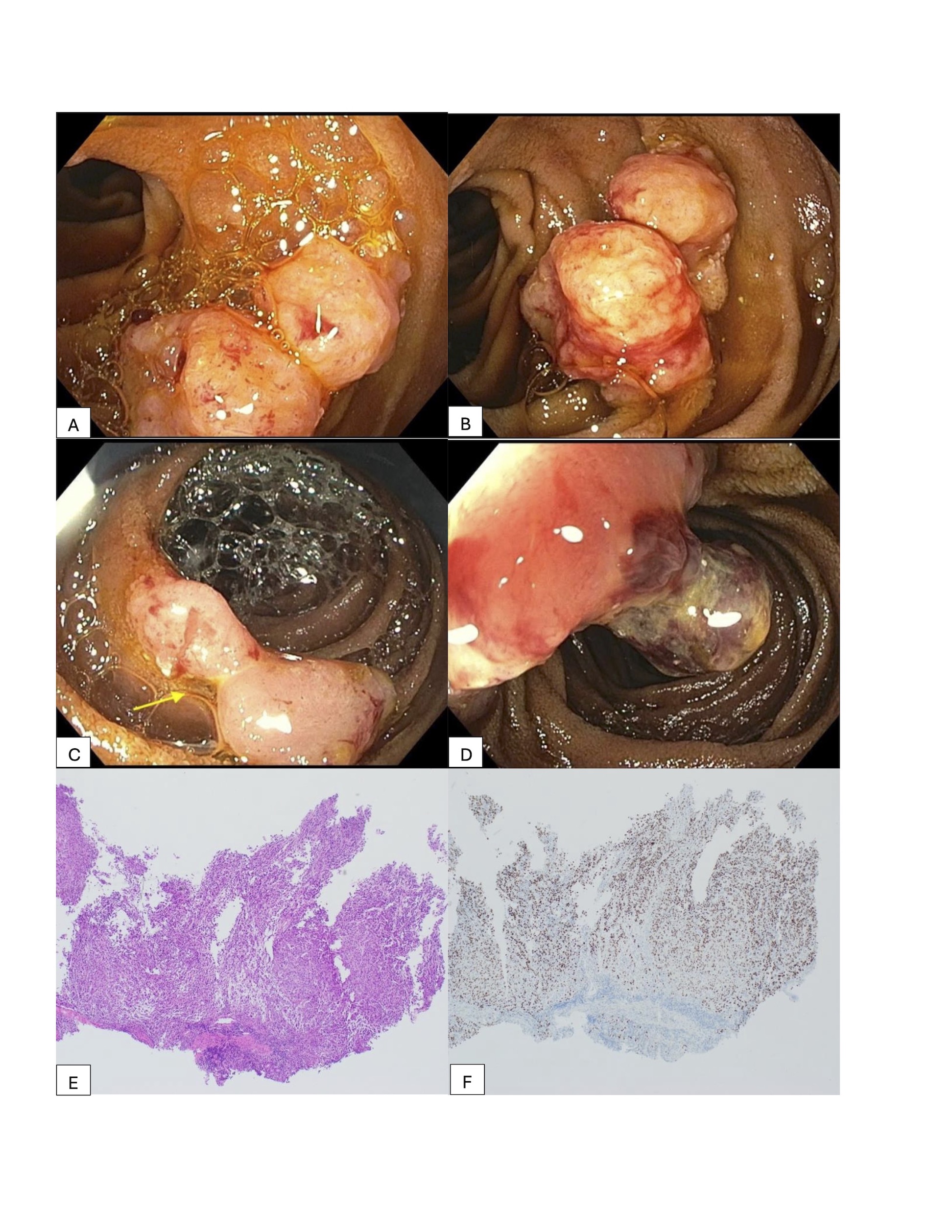
Figure: Figures A, B. Second portion of the duodenum showing metastatic mass from push enteroscopy. Figures C, D. Jejunum showing metastatic mass from push enteroscopy. Figure E. H&E stain of biopsy consisting predominantly of a dysplastic cellular infiltrate without identifiable small bowel mucosa. Figure F. PAX-8 stain of biopsy of the cellular infiltrate showing the expression of a transcription factor found in cells of renal origin.
Disclosures:
David Weinstein indicated no relevant financial relationships.
Jeremy Van indicated no relevant financial relationships.
Robert Toelke indicated no relevant financial relationships.
Mitchelle Zolotarevsky indicated no relevant financial relationships.
Michael Sprang indicated no relevant financial relationships.
David Weinstein, MD1, Jeremy Van, DO2, Robert Toelke, MD3, Mitchelle V. Zolotarevsky, MD4, Michael Sprang, MD3. P5002 - Renal Cell Carcinoma: An Unusual Presentation with Melena, ACG 2024 Annual Scientific Meeting Abstracts. Philadelphia, PA: American College of Gastroenterology.
