Tuesday Poster Session
Category: Small Intestine
P5014 - Endoscopic Omental (Graham) Patch and Duodenal Ulcer Repair Using a Helical Tack-Based System
Tuesday, October 29, 2024
10:30 AM - 4:00 PM ET
Location: Exhibit Hall E

Has Audio

Ronan Allencherril, MD
Houston Methodist
Houston, TX
Presenting Author(s)
Benjamin Warren, MD1, Ronan Allencherril, MD1, Thomas R. McCarty, MD, MPH2
1Houston Methodist Hospital, Houston, TX; 2Lynda K. and David M. Underwood Center for Digestive Disorders, Houston Methodist Hospital, Houston, TX
Introduction: A number of options are available for endoscopic closure of gastrointestinal defects including through-the-scope and over-the-scope clips as well as suturing devices. Here we describe a case of successful endoscopic omental (Graham) patch and perforated duodenal ulcer closure using a novel endoscopic helical tack-based system.
Case Description/Methods: A 59-year-old man presented to our emergency department with progressive abdominal pain and melena in the setting of chronic non-steroidal anti-inflammatory (NSAID) use. Computed tomography (CT) abdomen showed a perforation in the third portion of the duodenum with adjacent loculated phlegmon extending into the mid-abdomen and pelvis. He underwent exploratory laparotomy with omental (Graham) patch repair of a large, >2 cm perforated duodenal ulcer with clean edges and no signs of malignancy, suggestive of NSAID-associated ulceration. Evacuation of a retroperitoneal abscess was also performed and drain left in place. However, post-procedure the patient continued to have high volume bilious drain output ( >800 cc/day) with rising leukocytosis. Subsequent imaging showed persistent duodenal leak.
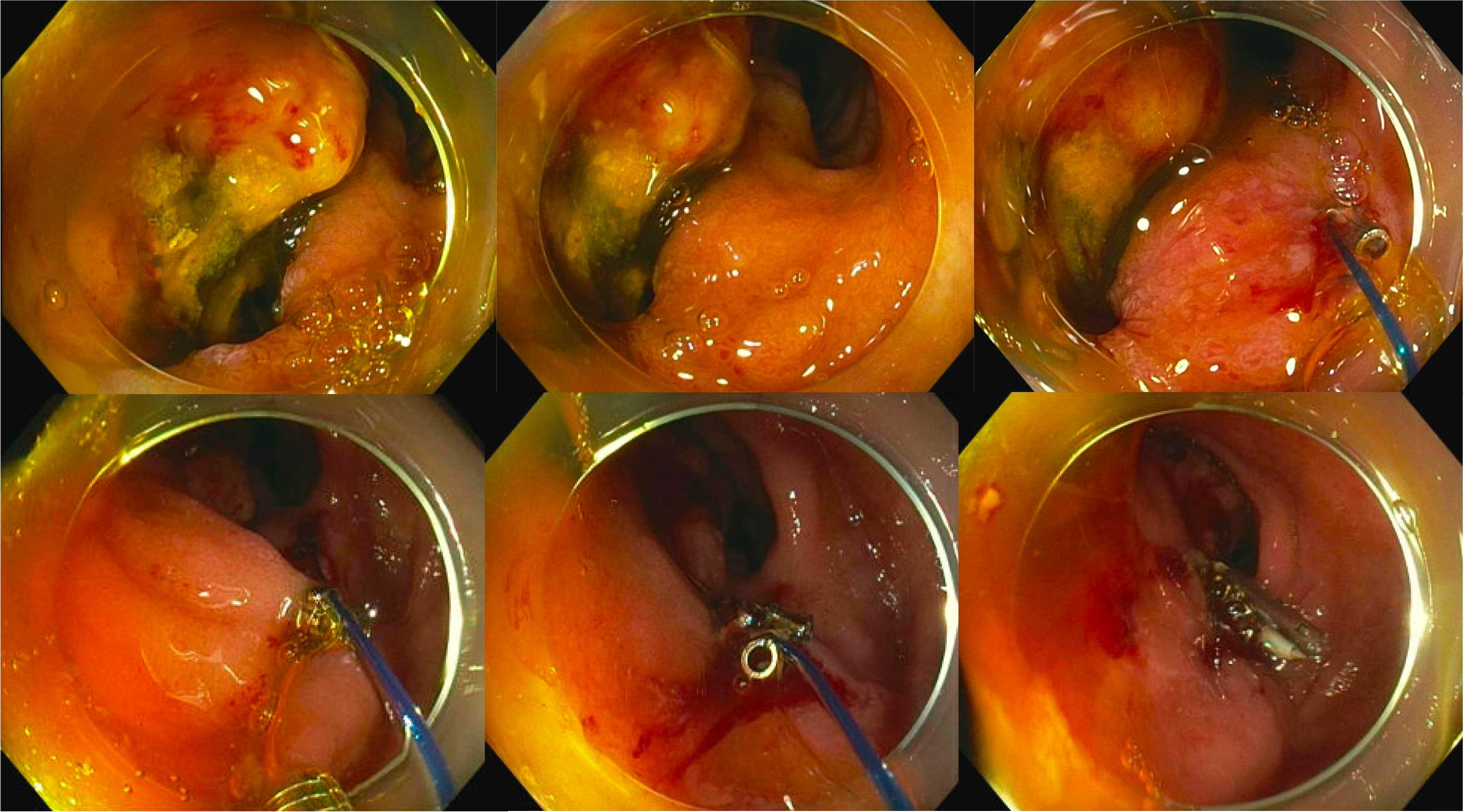
After discussion, the decision was made to proceed with endoscopy to evaluate for alternative leak sites and attempt endoscopic closure. Enteroscopy was then performed with distal attachment cap and noted a 15 mm perforation within the third portion of the duodenum. Mesentery was protruding through the full-thickness defect – Figure 1. Using a tack-based helical suturing device, four tacks were placed in a running fashion into the wall of the duodenum to appose the defect margins and cinched with excellent tissue approximation. Given full-thickness nature of the injury, two additional endoscopic hemoclips were placed to ensure closure. Under fluoroscopy, injection of luminal contrast revealed no extravasation, confirming successful closure. Drain output diminished rapidly the following day and diet was eventually advanced without issue. Post-procedure upper gastrointestinal contrast series (UGIs) revealed mild narrowing at the third portion of the duodenum with no contrast extravasation – confirming successful endoscopic omental (Graham) patch and perforated duodenal ulcer closure.
Discussion: This case highlights a novel approach to achieving endoscopic full-thickness closure of a perforated duodenal ulcer. Selecting the optimal modality depends on a variety of factors, including defect size and location within the gastrointestinal tract.

Disclosures:
Benjamin Warren, MD1, Ronan Allencherril, MD1, Thomas R. McCarty, MD, MPH2. P5014 - Endoscopic Omental (Graham) Patch and Duodenal Ulcer Repair Using a Helical Tack-Based System, ACG 2024 Annual Scientific Meeting Abstracts. Philadelphia, PA: American College of Gastroenterology.
1Houston Methodist Hospital, Houston, TX; 2Lynda K. and David M. Underwood Center for Digestive Disorders, Houston Methodist Hospital, Houston, TX
Introduction: A number of options are available for endoscopic closure of gastrointestinal defects including through-the-scope and over-the-scope clips as well as suturing devices. Here we describe a case of successful endoscopic omental (Graham) patch and perforated duodenal ulcer closure using a novel endoscopic helical tack-based system.
Case Description/Methods: A 59-year-old man presented to our emergency department with progressive abdominal pain and melena in the setting of chronic non-steroidal anti-inflammatory (NSAID) use. Computed tomography (CT) abdomen showed a perforation in the third portion of the duodenum with adjacent loculated phlegmon extending into the mid-abdomen and pelvis. He underwent exploratory laparotomy with omental (Graham) patch repair of a large, >2 cm perforated duodenal ulcer with clean edges and no signs of malignancy, suggestive of NSAID-associated ulceration. Evacuation of a retroperitoneal abscess was also performed and drain left in place. However, post-procedure the patient continued to have high volume bilious drain output ( >800 cc/day) with rising leukocytosis. Subsequent imaging showed persistent duodenal leak.
After discussion, the decision was made to proceed with endoscopy to evaluate for alternative leak sites and attempt endoscopic closure. Enteroscopy was then performed with distal attachment cap and noted a 15 mm perforation within the third portion of the duodenum. Mesentery was protruding through the full-thickness defect – Figure 1. Using a tack-based helical suturing device, four tacks were placed in a running fashion into the wall of the duodenum to appose the defect margins and cinched with excellent tissue approximation. Given full-thickness nature of the injury, two additional endoscopic hemoclips were placed to ensure closure. Under fluoroscopy, injection of luminal contrast revealed no extravasation, confirming successful closure. Drain output diminished rapidly the following day and diet was eventually advanced without issue. Post-procedure upper gastrointestinal contrast series (UGIs) revealed mild narrowing at the third portion of the duodenum with no contrast extravasation – confirming successful endoscopic omental (Graham) patch and perforated duodenal ulcer closure.
Discussion: This case highlights a novel approach to achieving endoscopic full-thickness closure of a perforated duodenal ulcer. Selecting the optimal modality depends on a variety of factors, including defect size and location within the gastrointestinal tract.

Figure: Full-thickness ulceration in the third portion of the duodenum and successful endoscopic closure using a novel endoscopic helical tack-based system
Disclosures:
Benjamin Warren indicated no relevant financial relationships.
Ronan Allencherril indicated no relevant financial relationships.
Thomas McCarty indicated no relevant financial relationships.
Benjamin Warren, MD1, Ronan Allencherril, MD1, Thomas R. McCarty, MD, MPH2. P5014 - Endoscopic Omental (Graham) Patch and Duodenal Ulcer Repair Using a Helical Tack-Based System, ACG 2024 Annual Scientific Meeting Abstracts. Philadelphia, PA: American College of Gastroenterology.
