Tuesday Poster Session
Category: Stomach
P5090 - Renal Cell Carcinoma's Stomach Detour: An Unusual Metastasis
Tuesday, October 29, 2024
10:30 AM - 4:00 PM ET
Location: Exhibit Hall E

Has Audio

Daniel Bleykhman, MD
Mount Sinai South Nassau, Icahn School of Medicine at Mount Sinai
Oceanside, NY
Presenting Author(s)
Daniel Bleykhman, MD1, Samuel Huang, MD1, Neal Shah, DO1, Jonathan Vincent M. Reyes, MD, MHA2, Pranay Srivastava, MD1, Rashmi Advani, MD1
1Mount Sinai South Nassau, Icahn School of Medicine at Mount Sinai, Oceanside, NY; 2Mount Sinai South Nassau, Oceanside, NY
Introduction: Renal Cell Carcinoma (RCC) metastasis to the stomach is an exceptionally rare event. Despite known dependability of clinical and CT diagnostic tools, immunohistochemistry staining for transcription factors of the normal renal parenchyma is increasingly being used. We present a case report of an 81-year-old female found to have a rare metastasis of RCC to the stomach, confirmed by biopsy and immunohistochemistry. This case report highlights the diagnostic challenges and complexities in patients with surprising sources of gastroenterological masses.
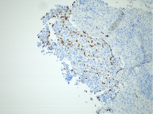
Case Description/Methods: The patient was an 81-year-old woman with history of Lynch syndrome, breast cancer, colon cancer and metastatic renal cell carcinoma diagnosed in November 2021. Patient presented with worsening back pain, weight loss, and low appetite. CT of the lumbar spine showed a new mass in the region of the gastric fundus, later confirmed on MRI lumbar spine and PET scan. Given her Lynch syndrome history, differential diagnosis included a primary gastric malignancy. Concomitant T12 mass was biopsied and found to be positive for metastasis from clear cell renal carcinoma. An EGD was performed showing a large, submucosal lesion with a central depression and ulcerated/necrotic core in the gastric fundus. Biopsies were taken showing metastatic carcinoma. Further pathology results revealed tumor cells staining positively with PAX8. Comparison to previous lytic lesion biopsy showed a similar morphology suggesting a diagnosis of metastatic renal cell carcinoma.
Discussion: The association of Renal Cell Carcinoma (RCC) and metastasis to the stomach is exceptionally rare. Gastric metastasis from RCC appears as an ulcer or tumor that is invading the submucosal layer of the upper or middle bodies of the stomach. This case was unusual in that the chief complaint was back pain, and initial surveillance involved a Spinal MRI that only then revealed the gastric fundal mass. An EGD with biopsy allows for definitive diagnosis and understanding of the tumor origin. The use of PAX8 and PAX2 immunohistochemical staining has become a reliable method for testing primary and metastatic renal neoplasm tissue. A more conservative treatment approach was chosen, as the limited data we have has not shown significant benefit to going through with surgery or endoscopic intervention. The patient is currently on chemotherapy regimen and has also received radiation therapy for coverage of other cancer sites.

Disclosures:
Daniel Bleykhman, MD1, Samuel Huang, MD1, Neal Shah, DO1, Jonathan Vincent M. Reyes, MD, MHA2, Pranay Srivastava, MD1, Rashmi Advani, MD1. P5090 - Renal Cell Carcinoma's Stomach Detour: An Unusual Metastasis, ACG 2024 Annual Scientific Meeting Abstracts. Philadelphia, PA: American College of Gastroenterology.
1Mount Sinai South Nassau, Icahn School of Medicine at Mount Sinai, Oceanside, NY; 2Mount Sinai South Nassau, Oceanside, NY
Introduction: Renal Cell Carcinoma (RCC) metastasis to the stomach is an exceptionally rare event. Despite known dependability of clinical and CT diagnostic tools, immunohistochemistry staining for transcription factors of the normal renal parenchyma is increasingly being used. We present a case report of an 81-year-old female found to have a rare metastasis of RCC to the stomach, confirmed by biopsy and immunohistochemistry. This case report highlights the diagnostic challenges and complexities in patients with surprising sources of gastroenterological masses.
Case Description/Methods: The patient was an 81-year-old woman with history of Lynch syndrome, breast cancer, colon cancer and metastatic renal cell carcinoma diagnosed in November 2021. Patient presented with worsening back pain, weight loss, and low appetite. CT of the lumbar spine showed a new mass in the region of the gastric fundus, later confirmed on MRI lumbar spine and PET scan. Given her Lynch syndrome history, differential diagnosis included a primary gastric malignancy. Concomitant T12 mass was biopsied and found to be positive for metastasis from clear cell renal carcinoma. An EGD was performed showing a large, submucosal lesion with a central depression and ulcerated/necrotic core in the gastric fundus. Biopsies were taken showing metastatic carcinoma. Further pathology results revealed tumor cells staining positively with PAX8. Comparison to previous lytic lesion biopsy showed a similar morphology suggesting a diagnosis of metastatic renal cell carcinoma.
Discussion: The association of Renal Cell Carcinoma (RCC) and metastasis to the stomach is exceptionally rare. Gastric metastasis from RCC appears as an ulcer or tumor that is invading the submucosal layer of the upper or middle bodies of the stomach. This case was unusual in that the chief complaint was back pain, and initial surveillance involved a Spinal MRI that only then revealed the gastric fundal mass. An EGD with biopsy allows for definitive diagnosis and understanding of the tumor origin. The use of PAX8 and PAX2 immunohistochemical staining has become a reliable method for testing primary and metastatic renal neoplasm tissue. A more conservative treatment approach was chosen, as the limited data we have has not shown significant benefit to going through with surgery or endoscopic intervention. The patient is currently on chemotherapy regimen and has also received radiation therapy for coverage of other cancer sites.

Figure: PAX-8 immunohistochemical stain demonstrates strong nuclear staining, an usual finding in gastrointestinal histology.
Disclosures:
Daniel Bleykhman indicated no relevant financial relationships.
Samuel Huang indicated no relevant financial relationships.
Neal Shah indicated no relevant financial relationships.
Jonathan Vincent Reyes indicated no relevant financial relationships.
Pranay Srivastava indicated no relevant financial relationships.
Rashmi Advani indicated no relevant financial relationships.
Daniel Bleykhman, MD1, Samuel Huang, MD1, Neal Shah, DO1, Jonathan Vincent M. Reyes, MD, MHA2, Pranay Srivastava, MD1, Rashmi Advani, MD1. P5090 - Renal Cell Carcinoma's Stomach Detour: An Unusual Metastasis, ACG 2024 Annual Scientific Meeting Abstracts. Philadelphia, PA: American College of Gastroenterology.
