Tuesday Poster Session
Category: Stomach
P5099 - Iron Pill Gastritis: An Under-Recognized Cause of Gastritis
Tuesday, October 29, 2024
10:30 AM - 4:00 PM ET
Location: Exhibit Hall E

Has Audio

Mena Saad, DO
Riverside University Health System
Moreno Valley, CA
Presenting Author(s)
Mena Saad, DO1, Matthew Orosa, DO1, Raul Hernandez, MD2, Sandra Saborio, BS3, Manish Shrestha, MD1, Jeremy Deisch, MD1
1Riverside University Health System, Moreno Valley, CA; 2Riverside University Health System, Redlands, CA; 3University of California Riverside School of Medicine, Riverside, CA
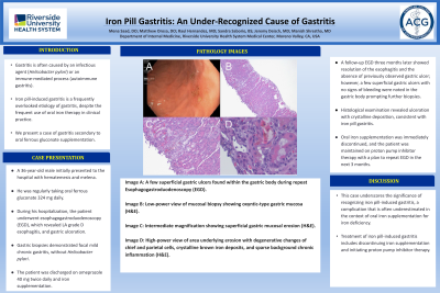
Introduction: Gastritis is often caused by an infectious agent (Helicobacter pylori) or an immune-mediated process (autoimmune gastritis). Iron pill-induced gastritis is a frequently overlooked etiology of gastritis, despite the frequent use of oral iron therapy in clinical practice. Here we present a case of iron pill-induced gastritis following ferrous gluconate supplementation.
Case Description/Methods: A 36-year-old male initially presented to the hospital with hematemesis and melena. He was regularly taking oral ferrous gluconate 324 mg daily following a diagnosis of low serum iron level on routine blood work. During his hospitalization, the patient underwent esophagogastroduodenoscopy (EGD), which revealed LA grade D esophagitis, and gastric ulceration. Gastric biopsies demonstrated focal mild chronic gastritis, without Helicobacter pylori. The patient was discharged on omeprazole 40 mg twice daily and iron supplementation. A follow-up EGD three months later showed resolution of the esophagitis and the absence of previously observed gastric ulcer. However, a few superficial gastric ulcers with no signs of bleeding were noted in the gastric body, prompting further biopsies. Histological examination revealed ulceration with crystalline deposition, consistent with iron pill gastritis. Oral iron supplementation was immediately discontinued, and the patient was maintained on proton pump inhibitor therapy with the plan to repeat EGD in the next 3 months.
Discussion: This case underscores the significance of recognizing iron pill-induced gastritis, a complication that is often underestimated in the context of oral iron supplementation for treating iron deficiency.

Disclosures:
Mena Saad, DO1, Matthew Orosa, DO1, Raul Hernandez, MD2, Sandra Saborio, BS3, Manish Shrestha, MD1, Jeremy Deisch, MD1. P5099 - Iron Pill Gastritis: An Under-Recognized Cause of Gastritis, ACG 2024 Annual Scientific Meeting Abstracts. Philadelphia, PA: American College of Gastroenterology.
1Riverside University Health System, Moreno Valley, CA; 2Riverside University Health System, Redlands, CA; 3University of California Riverside School of Medicine, Riverside, CA
Introduction: Gastritis is often caused by an infectious agent (Helicobacter pylori) or an immune-mediated process (autoimmune gastritis). Iron pill-induced gastritis is a frequently overlooked etiology of gastritis, despite the frequent use of oral iron therapy in clinical practice. Here we present a case of iron pill-induced gastritis following ferrous gluconate supplementation.
Case Description/Methods: A 36-year-old male initially presented to the hospital with hematemesis and melena. He was regularly taking oral ferrous gluconate 324 mg daily following a diagnosis of low serum iron level on routine blood work. During his hospitalization, the patient underwent esophagogastroduodenoscopy (EGD), which revealed LA grade D esophagitis, and gastric ulceration. Gastric biopsies demonstrated focal mild chronic gastritis, without Helicobacter pylori. The patient was discharged on omeprazole 40 mg twice daily and iron supplementation. A follow-up EGD three months later showed resolution of the esophagitis and the absence of previously observed gastric ulcer. However, a few superficial gastric ulcers with no signs of bleeding were noted in the gastric body, prompting further biopsies. Histological examination revealed ulceration with crystalline deposition, consistent with iron pill gastritis. Oral iron supplementation was immediately discontinued, and the patient was maintained on proton pump inhibitor therapy with the plan to repeat EGD in the next 3 months.
Discussion: This case underscores the significance of recognizing iron pill-induced gastritis, a complication that is often underestimated in the context of oral iron supplementation for treating iron deficiency.

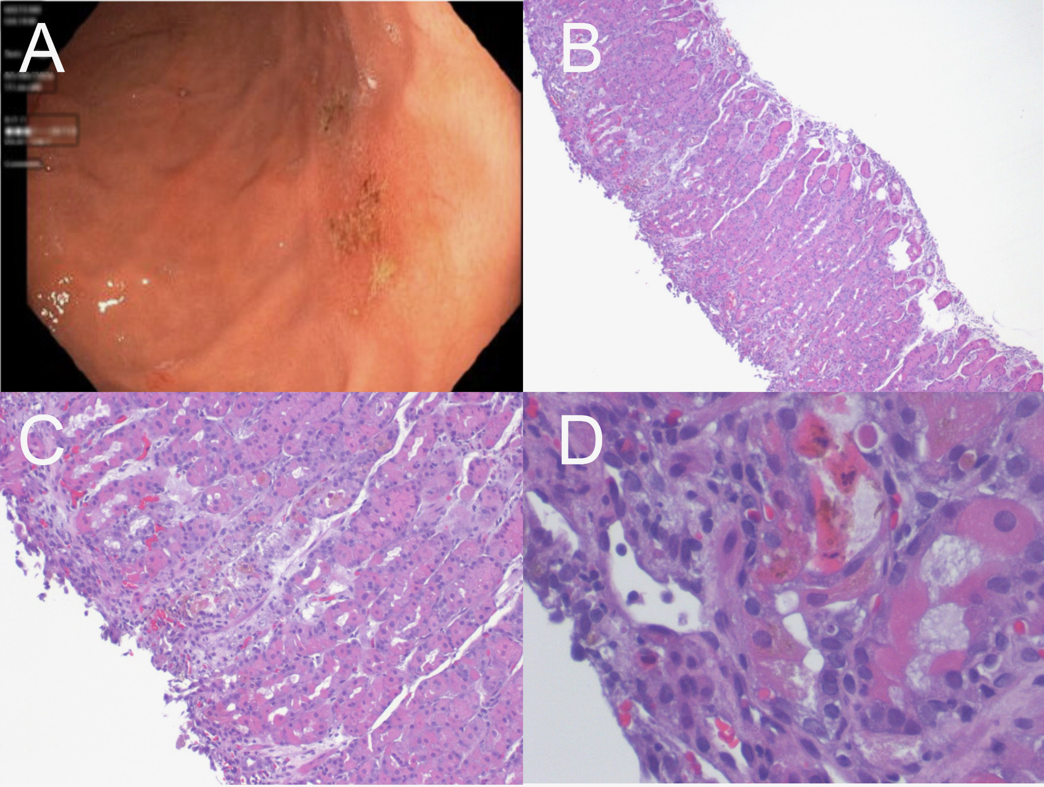
Figure: Image A: A few superficial gastric ulcers found within the gastric body during repeat Esophagogastroduodenoscopy (EGD).
Image B,C,D: Gastric mucosal histopathology demonstrating ulceration and brown crystalline iron deposition. Magnification in B is 100X, magnification in C is 200X, and magnification in D is 400X.
Image B,C,D: Gastric mucosal histopathology demonstrating ulceration and brown crystalline iron deposition. Magnification in B is 100X, magnification in C is 200X, and magnification in D is 400X.
Disclosures:
Mena Saad indicated no relevant financial relationships.
Matthew Orosa indicated no relevant financial relationships.
Raul Hernandez indicated no relevant financial relationships.
Sandra Saborio indicated no relevant financial relationships.
Manish Shrestha indicated no relevant financial relationships.
Jeremy Deisch indicated no relevant financial relationships.
Mena Saad, DO1, Matthew Orosa, DO1, Raul Hernandez, MD2, Sandra Saborio, BS3, Manish Shrestha, MD1, Jeremy Deisch, MD1. P5099 - Iron Pill Gastritis: An Under-Recognized Cause of Gastritis, ACG 2024 Annual Scientific Meeting Abstracts. Philadelphia, PA: American College of Gastroenterology.
