Tuesday Poster Session
Category: Stomach
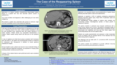
P5107 - A Case of Reappearance of the Long Lost Spleen
Tuesday, October 29, 2024
10:30 AM - 4:00 PM ET
Location: Exhibit Hall E

Has Audio

Justeena Joseph, MD
Long Island Community Hospital - NYU Langone Health System
New Hyde Park, NY
Presenting Author(s)
Justeena Joseph, MD1, Jiya Mulayamkuzhiyil, MD2, Jasbir Singh, MD3, Ivan Kharchenko, MD4, Nadia Khan, MBBS5, Papiya Begum, MD3, Zubin Tharayil, MD3, Prakash Viswanathan, MD, FACG3
1Long Island Community Hospital - NYU Langone Health System, New Hyde Park, NY; 2Long Island Community Hospital - NYU Langone Health System, Middle Island, NY; 3Long Island Community Hospital - NYU Langone Health System, Patchogue, NY; 4Long Island Community Hospital - NYU Langone Health System, Bellport, NY; 5Fatima Jinnah Medical University, Holbrook, NY
Introduction: Splenosis is a benign condition characterized by disseminated splenic tissue in a patient who has had traumatic splenic rupture or splenectomy. It is a rare condition and diagnosis is often challenging as it can mimic malignancy. We present a patient who presented with pyrexia of unknown origin and was incidentally found to have abdominal splenosis.
Case Description/Methods: A 54-year-old male with past medical history of splenectomy done at the age of 12 years presented to the ED with sepsis in the setting of a recent tick-borne infection. Given asplenia and an initial blood picture consistent with hemolysis, the patient was prophylactically started on treatment for babesiosis, however, treatment was discontinued once the workup for tick-borne illnesses came back negative. During the hospital course, the patient was found to have an incidental finding of nodular splenosis throughout the abdomen on CT imaging. The patient had a protracted course of febrile illness with no clear source. A biopsy of the nodular lesions was performed which was consistent with splenosis.
Discussion: Splenosis is an acquired ectopic auto-transplantation of splenic tissue following traumatic splenic rupture or splenectomy. Splenosis is reported in 25% of patients undergoing splenectomy following traumatic splenic injury. About 65% of splenosis cases are abdominopelvic with most common locations being parietal peritoneum, mesentery, greater omentum, serosal surface of the small and large bowel, and diaphragmatic surface. The pathogenesis include spillage of splenic pulp contents into surrounding cavities, hematogenous spread, or tissue growth in response to hypoxia. Patients are usually asymptomatic and splenosis is often incidentally found on imaging, with an average interval of two decades from the initial inciting event to diagnosis. The diagnosis can be made with CT, conventional MRI, and ultrasound. Nuclear scintigraphic studies help in differentiating splenosis from other malignant conditions. Biopsy of splenic nodules is rarely performed. The splenic nodules are insufficient to provide sufficient immunity against overwhelming post-splenectomy infections. The splenic tissue is non-invasive and does not warrant surgical removal unless complications ensue. Differential diagnosis of splenosis include accessory spleen, peritoneal carcinomatosis, and abdominal lymphoma. Physicians should consider splenosis in the differential diagnosis of soft tissue abdominal nodules in a patient with prior history of splenectomy.

Disclosures:
Justeena Joseph, MD1, Jiya Mulayamkuzhiyil, MD2, Jasbir Singh, MD3, Ivan Kharchenko, MD4, Nadia Khan, MBBS5, Papiya Begum, MD3, Zubin Tharayil, MD3, Prakash Viswanathan, MD, FACG3. P5107 - A Case of Reappearance of the Long Lost Spleen, ACG 2024 Annual Scientific Meeting Abstracts. Philadelphia, PA: American College of Gastroenterology.
1Long Island Community Hospital - NYU Langone Health System, New Hyde Park, NY; 2Long Island Community Hospital - NYU Langone Health System, Middle Island, NY; 3Long Island Community Hospital - NYU Langone Health System, Patchogue, NY; 4Long Island Community Hospital - NYU Langone Health System, Bellport, NY; 5Fatima Jinnah Medical University, Holbrook, NY
Introduction: Splenosis is a benign condition characterized by disseminated splenic tissue in a patient who has had traumatic splenic rupture or splenectomy. It is a rare condition and diagnosis is often challenging as it can mimic malignancy. We present a patient who presented with pyrexia of unknown origin and was incidentally found to have abdominal splenosis.
Case Description/Methods: A 54-year-old male with past medical history of splenectomy done at the age of 12 years presented to the ED with sepsis in the setting of a recent tick-borne infection. Given asplenia and an initial blood picture consistent with hemolysis, the patient was prophylactically started on treatment for babesiosis, however, treatment was discontinued once the workup for tick-borne illnesses came back negative. During the hospital course, the patient was found to have an incidental finding of nodular splenosis throughout the abdomen on CT imaging. The patient had a protracted course of febrile illness with no clear source. A biopsy of the nodular lesions was performed which was consistent with splenosis.
Discussion: Splenosis is an acquired ectopic auto-transplantation of splenic tissue following traumatic splenic rupture or splenectomy. Splenosis is reported in 25% of patients undergoing splenectomy following traumatic splenic injury. About 65% of splenosis cases are abdominopelvic with most common locations being parietal peritoneum, mesentery, greater omentum, serosal surface of the small and large bowel, and diaphragmatic surface. The pathogenesis include spillage of splenic pulp contents into surrounding cavities, hematogenous spread, or tissue growth in response to hypoxia. Patients are usually asymptomatic and splenosis is often incidentally found on imaging, with an average interval of two decades from the initial inciting event to diagnosis. The diagnosis can be made with CT, conventional MRI, and ultrasound. Nuclear scintigraphic studies help in differentiating splenosis from other malignant conditions. Biopsy of splenic nodules is rarely performed. The splenic nodules are insufficient to provide sufficient immunity against overwhelming post-splenectomy infections. The splenic tissue is non-invasive and does not warrant surgical removal unless complications ensue. Differential diagnosis of splenosis include accessory spleen, peritoneal carcinomatosis, and abdominal lymphoma. Physicians should consider splenosis in the differential diagnosis of soft tissue abdominal nodules in a patient with prior history of splenectomy.

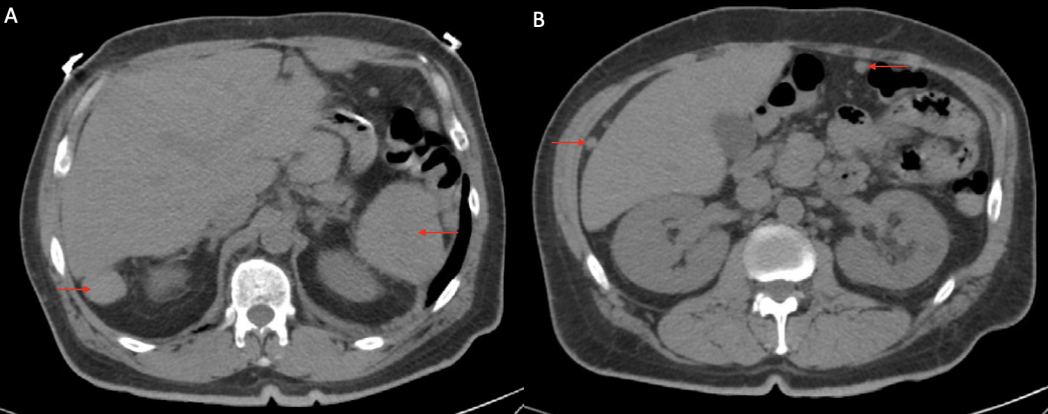
Figure: A: CT abdomen/pelvis showing larger nodules (red arrows) representing disseminated splenosis
B: CT abdomen/pelvis demonstrating smaller nodules (red arrows) of disseminated splenosis
B: CT abdomen/pelvis demonstrating smaller nodules (red arrows) of disseminated splenosis
Disclosures:
Justeena Joseph indicated no relevant financial relationships.
Jiya Mulayamkuzhiyil indicated no relevant financial relationships.
Jasbir Singh indicated no relevant financial relationships.
Ivan Kharchenko indicated no relevant financial relationships.
Nadia Khan indicated no relevant financial relationships.
Papiya Begum indicated no relevant financial relationships.
Zubin Tharayil indicated no relevant financial relationships.
Prakash Viswanathan indicated no relevant financial relationships.
Justeena Joseph, MD1, Jiya Mulayamkuzhiyil, MD2, Jasbir Singh, MD3, Ivan Kharchenko, MD4, Nadia Khan, MBBS5, Papiya Begum, MD3, Zubin Tharayil, MD3, Prakash Viswanathan, MD, FACG3. P5107 - A Case of Reappearance of the Long Lost Spleen, ACG 2024 Annual Scientific Meeting Abstracts. Philadelphia, PA: American College of Gastroenterology.
