Tuesday Poster Session
Category: Stomach
P5124 - Gastrointestinal Stromal Tumor Presenting at Gastroesophageal Junction: Case Report and Diagnostic Implications
Tuesday, October 29, 2024
10:30 AM - 4:00 PM ET
Location: Exhibit Hall E

Has Audio

Aparna Macharla, MBBS
Kasturba Medical College, Manipal
Bangalore, Karnataka, India
Presenting Author(s)
Muskan Jain, MBBS1, Vatsalya Choudhary, MBBS2, Jegan Mohan S, MBBS, MS3,Nithin Davuluri, MBBS4, Aparna Macharla, MBBS5
1Kasturba Medical College of Manipal, Central Delhi, Delhi, India; 2Kasturba Medical College of Manipal, Dhanbad, Jharkhand, India; 3Surgical Gastroenterology, Manipal, Karnataka, India; 4Kasturba Medical College, Manipal, Raichur, Karnataka, India; 5Kasturba Medical College, Manipal, Bangalore, Karnataka, India
Introduction: Gastrointestinal Stromal Tumor(GIST) is one of the rare causes of gastrointestinal tumors,accounting for 1-3% of all GI malignancies. Most commonly arise from the stomach(60%) but can also occur from the small bowel, esophagus,or colon.Presentation varies from incidental findings during endoscopies to fatal GI bleeding or bowel obstruction.Due to the malignant potential associated with extra-intestinal GIST,they are almost essential to diagnose.We report a GIST presenting at the gastroesophageal(GE) junction treated with Combined Endo-Laparoscopic Surgery.
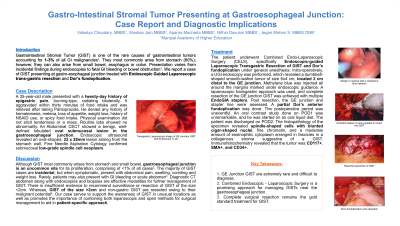
Case Description/Methods: A 35-year-old male presented with a twenty-day history of epigastric pain, burning-type, radiating bilaterally.It aggravated within thirty minutes of food intake and relieved after taking Pantoprazole.He denied any history of hematemesis,melena,weight loss or history of NSAID use.Physical examination did not elicit tenderness or a mass.Routine labs showed no abnormality.An Abdominal CECT showed a benign, lobulated oval submucosal lesion in the GE junction.EUS revealed an oval-shaped,22x28mm lesion arising from the stomach wall.FNAC confirmed submucosal spindle cell neoplasm.The patient underwent Endoscopic-guided Laparoscopic Transgastric Resection of GIST and Dor fundoplication.Intra-operatively, a UGI endoscopy revealed a dumbbell-shaped smooth-walled tumor of size 6x4 cm,< 2 cm proximal to the GE junction.Complete resection of GE junction GIST was done with partial Dor anterior fundoplication.The HPE of the specimen revealed spindle-shaped cells with blunted cigar-shaped nuclei and a moderate amount of eosinophilic cytoplasm arranged in fascicles in a collagenous stroma suggestive of a GIST.IHC revealed that the tumor was CD117+,SMA+,and CD34+.
Discussion: Although GIST most commonly arises from the stomach and small bowel, the GE junction is an uncommon site for its proliferation,comprising < 1% of all cases.Most cases are incidental, but symptomatic cases can present with abdominal pain,swelling and vomiting.Rarely, patients may also present with GI bleeding or acute abdomen.Diagnostic CT and endoscopies are effective modalities for further management of GIST.There is insufficient evidence for surveillance or resection of GIST< 2cm.GIST >2cm and non-gastric GIST are resected owing to their malignant potential.Our case supports the awareness of GIST in unusual locations.It promotes the importance of combining both laparoscopic and open methods for surgical management to aid in a patient-specific approach.

Disclosures:
Muskan Jain, MBBS1, Vatsalya Choudhary, MBBS2, Jegan Mohan S, MBBS, MS3,Nithin Davuluri, MBBS4, Aparna Macharla, MBBS5. P5124 - Gastrointestinal Stromal Tumor Presenting at Gastroesophageal Junction: Case Report and Diagnostic Implications, ACG 2024 Annual Scientific Meeting Abstracts. Philadelphia, PA: American College of Gastroenterology.
1Kasturba Medical College of Manipal, Central Delhi, Delhi, India; 2Kasturba Medical College of Manipal, Dhanbad, Jharkhand, India; 3Surgical Gastroenterology, Manipal, Karnataka, India; 4Kasturba Medical College, Manipal, Raichur, Karnataka, India; 5Kasturba Medical College, Manipal, Bangalore, Karnataka, India
Introduction: Gastrointestinal Stromal Tumor(GIST) is one of the rare causes of gastrointestinal tumors,accounting for 1-3% of all GI malignancies. Most commonly arise from the stomach(60%) but can also occur from the small bowel, esophagus,or colon.Presentation varies from incidental findings during endoscopies to fatal GI bleeding or bowel obstruction.Due to the malignant potential associated with extra-intestinal GIST,they are almost essential to diagnose.We report a GIST presenting at the gastroesophageal(GE) junction treated with Combined Endo-Laparoscopic Surgery.
Case Description/Methods: A 35-year-old male presented with a twenty-day history of epigastric pain, burning-type, radiating bilaterally.It aggravated within thirty minutes of food intake and relieved after taking Pantoprazole.He denied any history of hematemesis,melena,weight loss or history of NSAID use.Physical examination did not elicit tenderness or a mass.Routine labs showed no abnormality.An Abdominal CECT showed a benign, lobulated oval submucosal lesion in the GE junction.EUS revealed an oval-shaped,22x28mm lesion arising from the stomach wall.FNAC confirmed submucosal spindle cell neoplasm.The patient underwent Endoscopic-guided Laparoscopic Transgastric Resection of GIST and Dor fundoplication.Intra-operatively, a UGI endoscopy revealed a dumbbell-shaped smooth-walled tumor of size 6x4 cm,< 2 cm proximal to the GE junction.Complete resection of GE junction GIST was done with partial Dor anterior fundoplication.The HPE of the specimen revealed spindle-shaped cells with blunted cigar-shaped nuclei and a moderate amount of eosinophilic cytoplasm arranged in fascicles in a collagenous stroma suggestive of a GIST.IHC revealed that the tumor was CD117+,SMA+,and CD34+.
Discussion: Although GIST most commonly arises from the stomach and small bowel, the GE junction is an uncommon site for its proliferation,comprising < 1% of all cases.Most cases are incidental, but symptomatic cases can present with abdominal pain,swelling and vomiting.Rarely, patients may also present with GI bleeding or acute abdomen.Diagnostic CT and endoscopies are effective modalities for further management of GIST.There is insufficient evidence for surveillance or resection of GIST< 2cm.GIST >2cm and non-gastric GIST are resected owing to their malignant potential.Our case supports the awareness of GIST in unusual locations.It promotes the importance of combining both laparoscopic and open methods for surgical management to aid in a patient-specific approach.

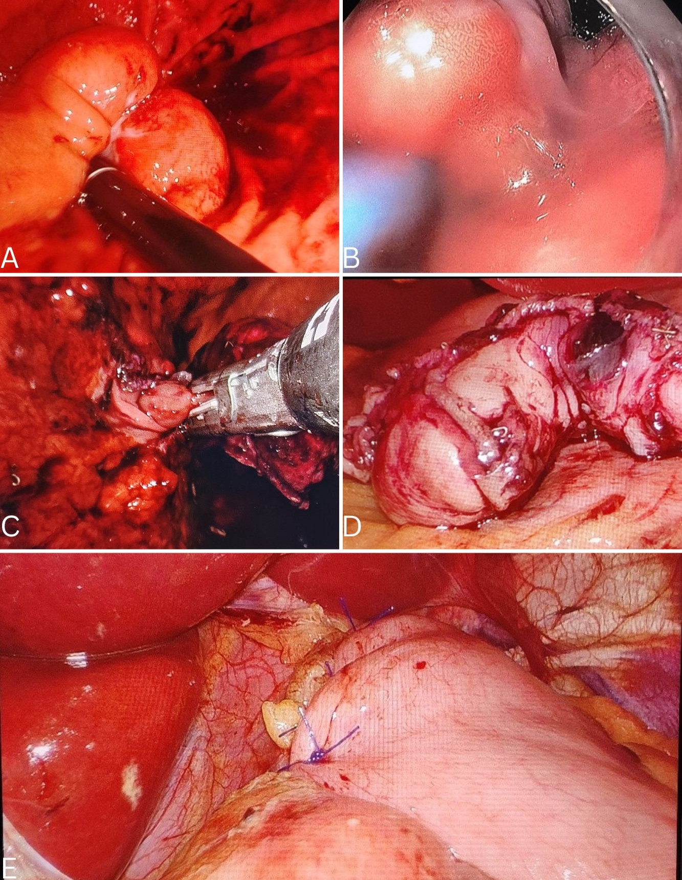
Figure: Figure A. Trans gastric Laparoscopic image of GE Junction GIST with Endoscopy in situ.
Figure B. Margin is marked endoscopically with a methylene blue injection.
Figure C. EndoGIA stapler is being applied to resect the GIST.
Figure D. Resected specimen of GIST.
Figure E. Dors fundoplication post-resection.
Figure B. Margin is marked endoscopically with a methylene blue injection.
Figure C. EndoGIA stapler is being applied to resect the GIST.
Figure D. Resected specimen of GIST.
Figure E. Dors fundoplication post-resection.
Disclosures:
Muskan Jain indicated no relevant financial relationships.
Vatsalya Choudhary indicated no relevant financial relationships.
Jegan Mohan S indicated no relevant financial relationships.
Nithin Davuluri indicated no relevant financial relationships.
Aparna Macharla indicated no relevant financial relationships.
Muskan Jain, MBBS1, Vatsalya Choudhary, MBBS2, Jegan Mohan S, MBBS, MS3,Nithin Davuluri, MBBS4, Aparna Macharla, MBBS5. P5124 - Gastrointestinal Stromal Tumor Presenting at Gastroesophageal Junction: Case Report and Diagnostic Implications, ACG 2024 Annual Scientific Meeting Abstracts. Philadelphia, PA: American College of Gastroenterology.

