Tuesday Poster Session
Category: Stomach
P5132 - A Rare Complication of Percutaneous Endoscopic Gastrostomy Tube Placement With Perforation of the Sigmoid Colon
Tuesday, October 29, 2024
10:30 AM - 4:00 PM ET
Location: Exhibit Hall E

Has Audio

Kwabena Asafo-Agyei, MBChB
Medical College of Georgia, Augusta University
Augusta, GA
Presenting Author(s)
Kwabena Asafo-Agyei, MBChB1, John Kirkikis, MD2, Dustin Hadley, PA-C2, Prince Tano. Djan, MD3
1Medical College of Georgia, Augusta University, Augusta, GA; 2GIS, Shreveport, LA; 3Cone Health Alamance, Burlington, NC
Introduction: Percutaneous endoscopic gastrostomy tube (PEG) tube placement is a widespread technique for enteral feeding to patients who cannot tolerate oral nutrition and require long-term nutrition.
Several techniques, including the “pull,” “push,” and “introducer” techniques, transillumination, and needle “safe tracking,” may be employed for safe and correct PEG placement. Despite these, safeguard measures are not always successful, and perforation as a complication occurs in about 0.5-1.8%. Mortality complications are less than 1%.
Case Description/Methods: An 88-year-old male with a past medical and surgical history, including hypertension, dementia, hernia repair, and a nursing home resident, was admitted for PEG tube insertion due to failure to thrive. The patient was recently admitted about a month ago for small bowel obstruction and was suspected to have ileus.
This resolved without surgical intervention.
The “pull,” transillumination, and needle-safe tracking techniques were employed to properly position the PEG tube placement.
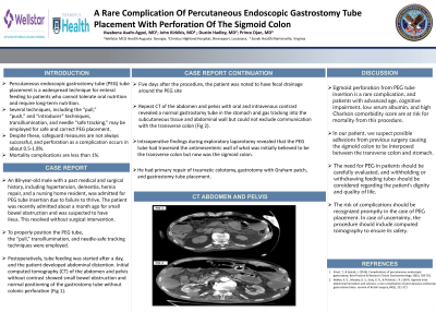
Postoperatively, tube feeding was started after a day, and the patient developed abdominal distention. Initial computed tomography (CT) of the abdomen and pelvis without contrast showed small bowel obstruction and normal positioning of the gastrostomy tube without colonic perforation (Fig 1).
5 days after the procedure, the patient was noted to have fecal drainage around the PEG site.
Repeat CT of the abdomen and pelvis with oral and intravenous contrast revealed a normal gastrostomy tube in the stomach and gas tracking into the subcutaneous tissue and abdominal wall but could not exclude communication with the transverse colon (Fig 2).
Intraoperative findings during exploratory laparotomy revealed the PEG tube had traversed the antimesenteric wall of what initially was believed to be the transverse colon but now was the sigmoid colon. He had primary repair of traumatic colotomy, gastrotomy with Graham patch, and gastrostomy tube placement.
Discussion: Sigmoid perforation from PEG tube insertion is a rare complication, and patients with advanced age, cognitive impairment, low serum albumin, and high Charlson comorbidity score are at risk for mortality from this procedure.
In our patient, we suspect possible adhesions from previous surgery causing the sigmoid colon to be interposed between the transverse colon and stomach. Gastroenterologists should carefully evaluate the need for PEG placement in these population groups and recognize complications promptly.

Disclosures:
Kwabena Asafo-Agyei, MBChB1, John Kirkikis, MD2, Dustin Hadley, PA-C2, Prince Tano. Djan, MD3. P5132 - A Rare Complication of Percutaneous Endoscopic Gastrostomy Tube Placement With Perforation of the Sigmoid Colon, ACG 2024 Annual Scientific Meeting Abstracts. Philadelphia, PA: American College of Gastroenterology.
1Medical College of Georgia, Augusta University, Augusta, GA; 2GIS, Shreveport, LA; 3Cone Health Alamance, Burlington, NC
Introduction: Percutaneous endoscopic gastrostomy tube (PEG) tube placement is a widespread technique for enteral feeding to patients who cannot tolerate oral nutrition and require long-term nutrition.
Several techniques, including the “pull,” “push,” and “introducer” techniques, transillumination, and needle “safe tracking,” may be employed for safe and correct PEG placement. Despite these, safeguard measures are not always successful, and perforation as a complication occurs in about 0.5-1.8%. Mortality complications are less than 1%.
Case Description/Methods: An 88-year-old male with a past medical and surgical history, including hypertension, dementia, hernia repair, and a nursing home resident, was admitted for PEG tube insertion due to failure to thrive. The patient was recently admitted about a month ago for small bowel obstruction and was suspected to have ileus.
This resolved without surgical intervention.
The “pull,” transillumination, and needle-safe tracking techniques were employed to properly position the PEG tube placement.
Postoperatively, tube feeding was started after a day, and the patient developed abdominal distention. Initial computed tomography (CT) of the abdomen and pelvis without contrast showed small bowel obstruction and normal positioning of the gastrostomy tube without colonic perforation (Fig 1).
5 days after the procedure, the patient was noted to have fecal drainage around the PEG site.
Repeat CT of the abdomen and pelvis with oral and intravenous contrast revealed a normal gastrostomy tube in the stomach and gas tracking into the subcutaneous tissue and abdominal wall but could not exclude communication with the transverse colon (Fig 2).
Intraoperative findings during exploratory laparotomy revealed the PEG tube had traversed the antimesenteric wall of what initially was believed to be the transverse colon but now was the sigmoid colon. He had primary repair of traumatic colotomy, gastrotomy with Graham patch, and gastrostomy tube placement.
Discussion: Sigmoid perforation from PEG tube insertion is a rare complication, and patients with advanced age, cognitive impairment, low serum albumin, and high Charlson comorbidity score are at risk for mortality from this procedure.
In our patient, we suspect possible adhesions from previous surgery causing the sigmoid colon to be interposed between the transverse colon and stomach. Gastroenterologists should carefully evaluate the need for PEG placement in these population groups and recognize complications promptly.

Figure: CT ABDOMEN AND PELVIS IMAGING WITH AND WITHOUT CONTRAST
Disclosures:
Kwabena Asafo-Agyei indicated no relevant financial relationships.
John Kirkikis indicated no relevant financial relationships.
Dustin Hadley indicated no relevant financial relationships.
Prince Djan indicated no relevant financial relationships.
Kwabena Asafo-Agyei, MBChB1, John Kirkikis, MD2, Dustin Hadley, PA-C2, Prince Tano. Djan, MD3. P5132 - A Rare Complication of Percutaneous Endoscopic Gastrostomy Tube Placement With Perforation of the Sigmoid Colon, ACG 2024 Annual Scientific Meeting Abstracts. Philadelphia, PA: American College of Gastroenterology.
