Tuesday Poster Session
Category: Stomach
P5135 - A Bizarre Case of a Bezoar - A Hairy Situation
Tuesday, October 29, 2024
10:30 AM - 4:00 PM ET
Location: Exhibit Hall E

Has Audio
- CW
Chelsea Wuthnow, MD, JD
Dallas Methodist
Flower Mound, TX
Presenting Author(s)
Chelsea Wuthnow, MD, JD1, Kyle Schneider, MD2, Armond Schwartz, MD3
1Dallas Methodist, Flower Mound, TX; 2Methodist Dallas Medical Center, Dallas, TX; 3Dallas Methodist, Dallas, TX
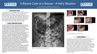
Introduction: A trichobezoar is an accumulation of hair and fibers within the gastrointestinal tract. They are most commonly found in children, typically with underlying psychiatric disorders. They can present with vague and unusual symptoms. We present a case of a 25 year old female with no psychiatric history and a chief complaint of lower extremity edema who was found to have a large trichobezoar missed on initial imaging.
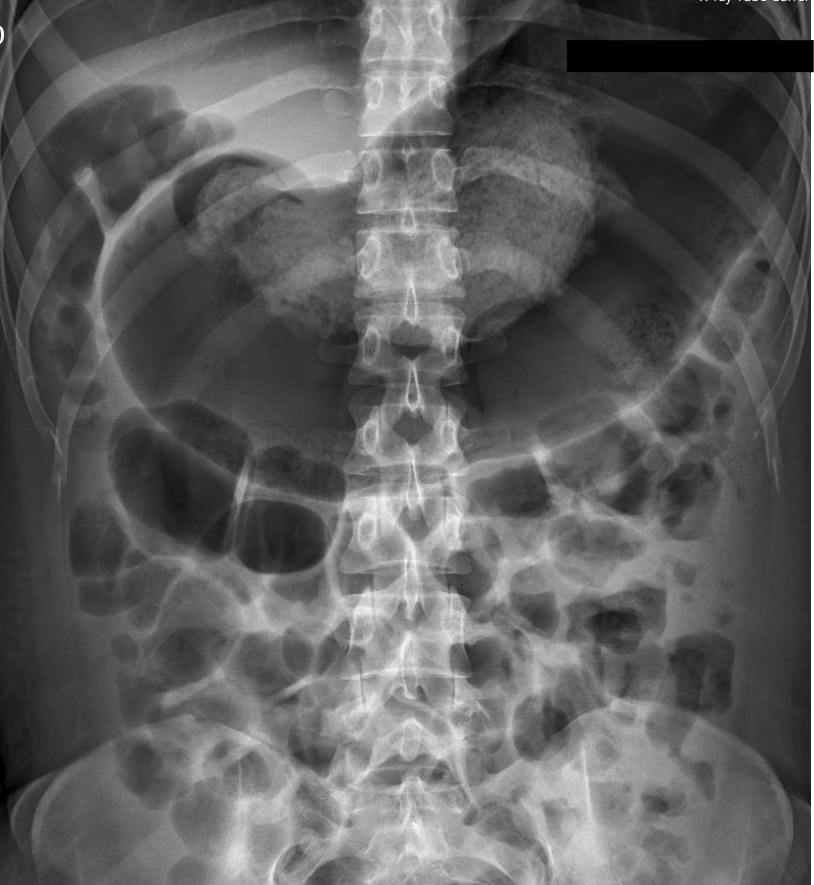
Case Description/Methods: A 25 year old female with no past medical history presented with leg swelling, intermittent abdominal discomfort and early satiety. Physical exam showed edema and a distended nontender abdomen with normal bowel sounds. Labs showed iron deficiency anemia and hypoalbuminemia with normal C reactive protein and negative celiac and anti nuclear antibodies. No proteinuria or thyroid abnormalities were found. X-ray abdomen was read with marked gastric distension. Computed tomography (CT) abdomen/pelvis with contrast was read as small bowel obstruction with transition point in the jejunum. Echocardiogram was unremarkable. CT enterography revealed foreign material in distal stomach extending into proximal duodenum with elongated solid appearing material within loops of jejunum. Retrospective view of the abdominal X-ray showed evidence of the bezoar. Upon further questioning patient admitted to eating her own hair and cloth fiber strands for several years. Esophagogastroduodenoscopy revealed a large trichobezoar in the gastric body extending into the second portion of the duodenum. Removal was not attempted due to composition and size of the bezoar. Total parental nutrition was given for several days and diet was slowly advanced. Patient tolerated a full liquid diet and discharged with surgical follow up.
Discussion: Trichobezoars make up 6% of all bezoars and are most often found in the stomach but can extend into the small intestine leading to “Rapunzel syndrome”. Large size and the chronic nature of such a bezoar can lead to a protein losing enteropathy as well as iron deficiency anemia. Pediatric literature has reported cases presenting with protein losing enteropathies, but review of adult literature reveals no such cases. Treatment options of such a bezoar can be limited as they are generally not amenable to chemical dissolution. Endoscopic removal can be attempted if small, however, if large, it is not recommended due to increased risk of complications. This case presents a challenging diagnostic and treatment dilemma for adult gastroenterologists

Disclosures:
Chelsea Wuthnow, MD, JD1, Kyle Schneider, MD2, Armond Schwartz, MD3. P5135 - A Bizarre Case of a Bezoar - A Hairy Situation, ACG 2024 Annual Scientific Meeting Abstracts. Philadelphia, PA: American College of Gastroenterology.
1Dallas Methodist, Flower Mound, TX; 2Methodist Dallas Medical Center, Dallas, TX; 3Dallas Methodist, Dallas, TX
Introduction: A trichobezoar is an accumulation of hair and fibers within the gastrointestinal tract. They are most commonly found in children, typically with underlying psychiatric disorders. They can present with vague and unusual symptoms. We present a case of a 25 year old female with no psychiatric history and a chief complaint of lower extremity edema who was found to have a large trichobezoar missed on initial imaging.
Case Description/Methods: A 25 year old female with no past medical history presented with leg swelling, intermittent abdominal discomfort and early satiety. Physical exam showed edema and a distended nontender abdomen with normal bowel sounds. Labs showed iron deficiency anemia and hypoalbuminemia with normal C reactive protein and negative celiac and anti nuclear antibodies. No proteinuria or thyroid abnormalities were found. X-ray abdomen was read with marked gastric distension. Computed tomography (CT) abdomen/pelvis with contrast was read as small bowel obstruction with transition point in the jejunum. Echocardiogram was unremarkable. CT enterography revealed foreign material in distal stomach extending into proximal duodenum with elongated solid appearing material within loops of jejunum. Retrospective view of the abdominal X-ray showed evidence of the bezoar. Upon further questioning patient admitted to eating her own hair and cloth fiber strands for several years. Esophagogastroduodenoscopy revealed a large trichobezoar in the gastric body extending into the second portion of the duodenum. Removal was not attempted due to composition and size of the bezoar. Total parental nutrition was given for several days and diet was slowly advanced. Patient tolerated a full liquid diet and discharged with surgical follow up.
Discussion: Trichobezoars make up 6% of all bezoars and are most often found in the stomach but can extend into the small intestine leading to “Rapunzel syndrome”. Large size and the chronic nature of such a bezoar can lead to a protein losing enteropathy as well as iron deficiency anemia. Pediatric literature has reported cases presenting with protein losing enteropathies, but review of adult literature reveals no such cases. Treatment options of such a bezoar can be limited as they are generally not amenable to chemical dissolution. Endoscopic removal can be attempted if small, however, if large, it is not recommended due to increased risk of complications. This case presents a challenging diagnostic and treatment dilemma for adult gastroenterologists

Figure: Initial Abdominal X-Ray
Disclosures:
Chelsea Wuthnow indicated no relevant financial relationships.
Kyle Schneider indicated no relevant financial relationships.
Armond Schwartz indicated no relevant financial relationships.
Chelsea Wuthnow, MD, JD1, Kyle Schneider, MD2, Armond Schwartz, MD3. P5135 - A Bizarre Case of a Bezoar - A Hairy Situation, ACG 2024 Annual Scientific Meeting Abstracts. Philadelphia, PA: American College of Gastroenterology.
