Tuesday Poster Session
Category: Liver
P4826 - An Interesting Case of Metastatic Hepatocellular Carcinoma Extending Into the Right Atrium
Tuesday, October 29, 2024
10:30 AM - 4:00 PM ET
Location: Exhibit Hall E

Has Audio

Kavya Priya Somu, MD
Centinela Hospital Medical Center
Los angeles, California
Presenting Author(s)
Saisree Reddy Adla Jala, MD1, Kavya Priya Somu, MD2, Nikitha Chellapuram, MD1, Lakshmi Deepika Gopalam, MD2, Varsha Reddy, MD2, Jonathan Hughes, 1, Mohsen Kheradpezhouh, MD2
1Centinela Hospital Medical Center, Los Angeles, CA; 2Centinela Hospital Medical Center, Inglewood, CA
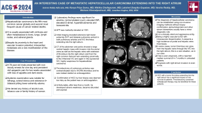
Introduction: Hepatocellular carcinoma is the fifth most common cancer globally. It is the second most frequent cause of cancer related deaths. It is usually associated with cirrhosis and often metastasizes to bone, lungs, lymph nodes, and adrenal glands. Despite its proximity to the heart and vascular invasion potential, intracardiac metastasis are a rare manifestation of the disease. Here we present an interesting case of metastatic hepatocellular carcinoma.
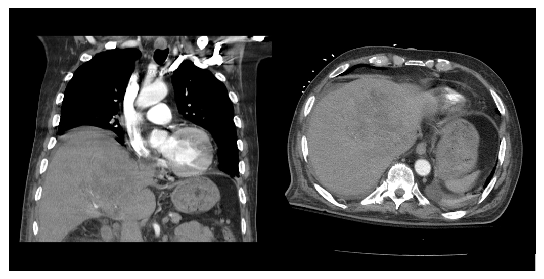
Case Description/Methods: A 78-year-old male presented with non-bloody emesis for one day and persistent dull epigastric pain for two weeks associated with loss of appetite and dark stools. Systemic examination was notable for lethargy, scleral icterus and extensive 4+ bilateral pitting lower extremity edema. He denied any history of alcohol use, tobacco use or family history of cancer. Laboratory findings were significant for anemia, normal platelet count, elevated INR, elevated D-dimer, hyperbilirubinemia and transaminitis. AFP was markedly elevated at 1361. Initial imaging revealed extensive right lower extremity DVT and bilateral pulmonary emboli in both pulmonary arteries and IVC thrombus extending into the right atrium. CT of the abdomen and pelvis showed a large central hepatic mass with invasion into the portal veins as well as the hepatic veins with extension of thrombus into the right atrium and from the right common femoral vein extending proximally to the infrarenal IVC and again in the suprarenal IVC, highly suspicious for hepatocellular carcinoma. Thrombectomy of pulmonary embolus was contraindicated due to IVC/RA thrombus and he was instead started on anticoagulation. Confirmation of HCC by liver biopsy was deemed too risky as the patient was on anticoagulation. Unfortunately, after less than a month, he developed severe weakness, became obtunded and expired.
Discussion: The diagnosis of hepatocellular carcinoma can be established using non-invasive imaging methods without biopsy confirmation; alpha-fetoprotein and other serum biomarkers usually have a minor diagnostic role. It is a clinically silent and aggressive entity. Being a highly vascular tumor with intravascular dissemination, it presents a high incidence of portal and hepatic veins thrombosis. In some cases, tumor thrombus can grow from the hepatic veins through the IVC into the right atrium; this is a rare situation, as in our case. It has a poor prognosis, with a reported median survival of 4–7 months in untreated patients. Prognosis with right atrium invasion is even worse.

Disclosures:
Saisree Reddy Adla Jala, MD1, Kavya Priya Somu, MD2, Nikitha Chellapuram, MD1, Lakshmi Deepika Gopalam, MD2, Varsha Reddy, MD2, Jonathan Hughes, 1, Mohsen Kheradpezhouh, MD2. P4826 - An Interesting Case of Metastatic Hepatocellular Carcinoma Extending Into the Right Atrium, ACG 2024 Annual Scientific Meeting Abstracts. Philadelphia, PA: American College of Gastroenterology.
1Centinela Hospital Medical Center, Los Angeles, CA; 2Centinela Hospital Medical Center, Inglewood, CA
Introduction: Hepatocellular carcinoma is the fifth most common cancer globally. It is the second most frequent cause of cancer related deaths. It is usually associated with cirrhosis and often metastasizes to bone, lungs, lymph nodes, and adrenal glands. Despite its proximity to the heart and vascular invasion potential, intracardiac metastasis are a rare manifestation of the disease. Here we present an interesting case of metastatic hepatocellular carcinoma.
Case Description/Methods: A 78-year-old male presented with non-bloody emesis for one day and persistent dull epigastric pain for two weeks associated with loss of appetite and dark stools. Systemic examination was notable for lethargy, scleral icterus and extensive 4+ bilateral pitting lower extremity edema. He denied any history of alcohol use, tobacco use or family history of cancer. Laboratory findings were significant for anemia, normal platelet count, elevated INR, elevated D-dimer, hyperbilirubinemia and transaminitis. AFP was markedly elevated at 1361. Initial imaging revealed extensive right lower extremity DVT and bilateral pulmonary emboli in both pulmonary arteries and IVC thrombus extending into the right atrium. CT of the abdomen and pelvis showed a large central hepatic mass with invasion into the portal veins as well as the hepatic veins with extension of thrombus into the right atrium and from the right common femoral vein extending proximally to the infrarenal IVC and again in the suprarenal IVC, highly suspicious for hepatocellular carcinoma. Thrombectomy of pulmonary embolus was contraindicated due to IVC/RA thrombus and he was instead started on anticoagulation. Confirmation of HCC by liver biopsy was deemed too risky as the patient was on anticoagulation. Unfortunately, after less than a month, he developed severe weakness, became obtunded and expired.
Discussion: The diagnosis of hepatocellular carcinoma can be established using non-invasive imaging methods without biopsy confirmation; alpha-fetoprotein and other serum biomarkers usually have a minor diagnostic role. It is a clinically silent and aggressive entity. Being a highly vascular tumor with intravascular dissemination, it presents a high incidence of portal and hepatic veins thrombosis. In some cases, tumor thrombus can grow from the hepatic veins through the IVC into the right atrium; this is a rare situation, as in our case. It has a poor prognosis, with a reported median survival of 4–7 months in untreated patients. Prognosis with right atrium invasion is even worse.

Figure: Coronal and axial CT abdomen showing large hepatic mass extending into the IVC and right atrium
Disclosures:
Saisree Reddy Adla Jala indicated no relevant financial relationships.
Kavya Priya Somu indicated no relevant financial relationships.
Nikitha Chellapuram indicated no relevant financial relationships.
Lakshmi Deepika Gopalam indicated no relevant financial relationships.
Varsha Reddy indicated no relevant financial relationships.
Jonathan Hughes indicated no relevant financial relationships.
Mohsen Kheradpezhouh indicated no relevant financial relationships.
Saisree Reddy Adla Jala, MD1, Kavya Priya Somu, MD2, Nikitha Chellapuram, MD1, Lakshmi Deepika Gopalam, MD2, Varsha Reddy, MD2, Jonathan Hughes, 1, Mohsen Kheradpezhouh, MD2. P4826 - An Interesting Case of Metastatic Hepatocellular Carcinoma Extending Into the Right Atrium, ACG 2024 Annual Scientific Meeting Abstracts. Philadelphia, PA: American College of Gastroenterology.
