Tuesday Poster Session
Category: IBD
P4424 - Symptomatic Mucocele Following Proctocolectomy With Ileal Pouch Anal Anastomosis for Ulcerative Colitis: A Case Report
Tuesday, October 29, 2024
10:30 AM - 4:00 PM ET
Location: Exhibit Hall E

Has Audio
- PR
Prajith Raj Ramesh, MBBS
Mayo Clinic
Rochester, MN
Presenting Author(s)
Prajith Raj Ramesh, MBBS, Amanda M. Johnson, MD
Mayo Clinic, Rochester, MN
Introduction: Total proctocolectomy with ileal pouch-anal anastomosis (IPAA) is a surgical option for refractory ulcerative colitis (UC). Potential complications include pouchitis, strictures, and bowel obstruction among others. Herein we report a rare complication following IPAA, a benign mucocele leading to obstruction.
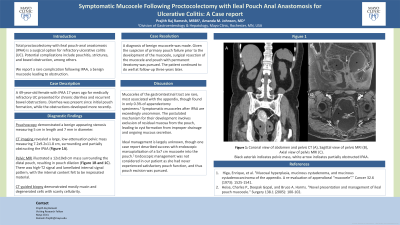
Case Description/Methods: A 49-year-old female with a history of IPAA 17 years ago for medically refractory UC presented for chronic diarrhea and recurrent bowel obstructions. Diarrhea was present since initial pouch formation, while the obstructions developed more recently. CT imaging revealed a large, low-attenuation pelvic mass measuring 7.2x9.2x11.8 cm, surrounding and partially obstructing the IPAA (Figure 1A). Pouchoscopy demonstrated a benign appearing stenosis measuring 5 cm in length and 7 mm in diameter. Pelvic MRI illustrated a 12x10x8 cm mass surrounding the distal pouch, resulting in pouch dilation (Figure 1B and 1C). There was high-T2 signal and lamellated internal signal pattern, with the internal content felt to be inspissated material. CT-guided biopsy was obtained and demonstrated mostly mucin and degenerated cells with scanty cellularity.
A diagnosis of benign mucocele was made. Given the suspicion of primary pouch failure prior to the development of the mucocele, surgical resection of the mucocele and pouch with permanent ileostomy was pursued. The patient continued to do well at follow-up three years later.
Discussion: Mucoceles of the gastrointestinal tract are rare, most associated with the appendix, though found in only 0.3% of appendectomy specimens.1 Symptomatic mucoceles after IPAA are exceedingly uncommon. The postulated mechanism for their development involves exclusion of residual mucosa from the pouch, leading to cyst formation from improper drainage and ongoing mucous secretion. Ideal management is largely unknown, though one case report described success with endoscopic marsupialization of a 5x7 cm mucocele into the pouch.2 Endoscopic management was not considered in our patient as she had never experienced satisfactory pouch function, and thus pouch excision was pursued.
References
1) Higa, Enrique, et al. "Mucosal hyperplasia, mucinous cystadenoma, and mucinous cystadenocarcinoma of the appendix. A re‐evaluation of appendiceal “mucocele”." Cancer 32.6 (1973): 1525-1541.
2) Heise, Charles P., Deepak Gopal, and Bruce A. Harms. "Novel presentation and management of ileal pouch mucocele." Surgery 138.1 (2005): 100-102.

Disclosures:
Prajith Raj Ramesh, MBBS, Amanda M. Johnson, MD. P4424 - Symptomatic Mucocele Following Proctocolectomy With Ileal Pouch Anal Anastomosis for Ulcerative Colitis: A Case Report, ACG 2024 Annual Scientific Meeting Abstracts. Philadelphia, PA: American College of Gastroenterology.
Mayo Clinic, Rochester, MN
Introduction: Total proctocolectomy with ileal pouch-anal anastomosis (IPAA) is a surgical option for refractory ulcerative colitis (UC). Potential complications include pouchitis, strictures, and bowel obstruction among others. Herein we report a rare complication following IPAA, a benign mucocele leading to obstruction.
Case Description/Methods: A 49-year-old female with a history of IPAA 17 years ago for medically refractory UC presented for chronic diarrhea and recurrent bowel obstructions. Diarrhea was present since initial pouch formation, while the obstructions developed more recently. CT imaging revealed a large, low-attenuation pelvic mass measuring 7.2x9.2x11.8 cm, surrounding and partially obstructing the IPAA (Figure 1A). Pouchoscopy demonstrated a benign appearing stenosis measuring 5 cm in length and 7 mm in diameter. Pelvic MRI illustrated a 12x10x8 cm mass surrounding the distal pouch, resulting in pouch dilation (Figure 1B and 1C). There was high-T2 signal and lamellated internal signal pattern, with the internal content felt to be inspissated material. CT-guided biopsy was obtained and demonstrated mostly mucin and degenerated cells with scanty cellularity.
A diagnosis of benign mucocele was made. Given the suspicion of primary pouch failure prior to the development of the mucocele, surgical resection of the mucocele and pouch with permanent ileostomy was pursued. The patient continued to do well at follow-up three years later.
Discussion: Mucoceles of the gastrointestinal tract are rare, most associated with the appendix, though found in only 0.3% of appendectomy specimens.1 Symptomatic mucoceles after IPAA are exceedingly uncommon. The postulated mechanism for their development involves exclusion of residual mucosa from the pouch, leading to cyst formation from improper drainage and ongoing mucous secretion. Ideal management is largely unknown, though one case report described success with endoscopic marsupialization of a 5x7 cm mucocele into the pouch.2 Endoscopic management was not considered in our patient as she had never experienced satisfactory pouch function, and thus pouch excision was pursued.
References
1) Higa, Enrique, et al. "Mucosal hyperplasia, mucinous cystadenoma, and mucinous cystadenocarcinoma of the appendix. A re‐evaluation of appendiceal “mucocele”." Cancer 32.6 (1973): 1525-1541.
2) Heise, Charles P., Deepak Gopal, and Bruce A. Harms. "Novel presentation and management of ileal pouch mucocele." Surgery 138.1 (2005): 100-102.

Figure: Figure 1: Coronal view of abdomen and pelvis CT (A), Sagittal view of pelvis MRI (B), Axial view of pelvis MRI (C). Black asterisk indicates pelvic mass, white arrow indicates partially obstructed IPAA.
Disclosures:
Prajith Raj Ramesh indicated no relevant financial relationships.
Amanda Johnson indicated no relevant financial relationships.
Prajith Raj Ramesh, MBBS, Amanda M. Johnson, MD. P4424 - Symptomatic Mucocele Following Proctocolectomy With Ileal Pouch Anal Anastomosis for Ulcerative Colitis: A Case Report, ACG 2024 Annual Scientific Meeting Abstracts. Philadelphia, PA: American College of Gastroenterology.
